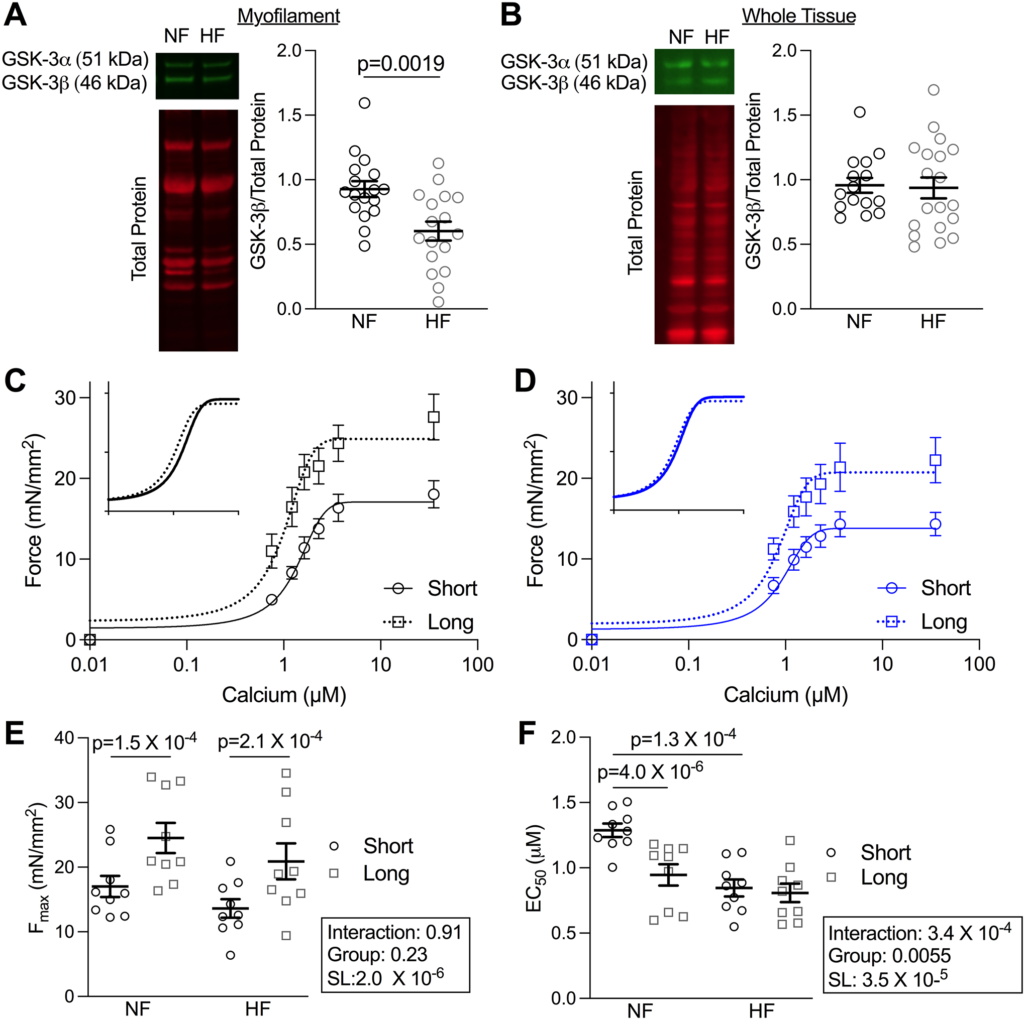
Figure 6. Myofilament-localized GSK-3β and LDA is reduced in human heart failure.

(A) Example blots and quantification of myofilament-enriched (n=17, p=0.0019 by unpaired t-test) and (B) Whole tissue preparation (n values: NF=15, HF=19) of LV from human NF and HF samples. (C) Mean force as a function of calcium concentration and fitted curves for skinned myocytes from the LV of NF and (D) HF patients from which measurements were taken at short (1.9 μm) and long (2.3 μm) SL. (E) Fmax and (F) EC50 depicted as mean± SEM (n = 9 myocytes from 3 patients). P-values were calculated from two-way repeated measures ANOVA with pair-wise comparisons.
