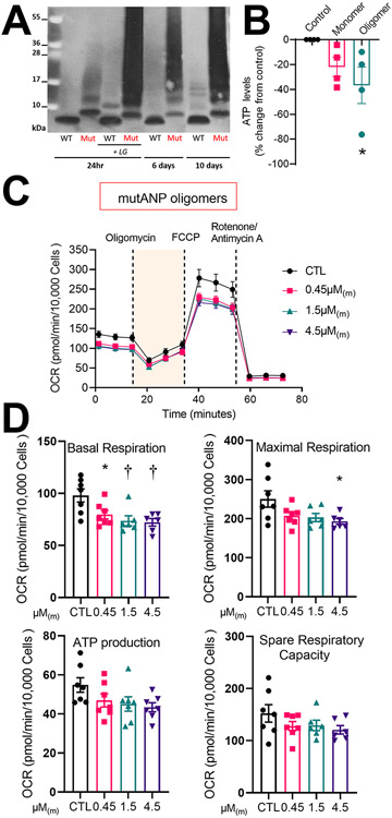
Figure 1. Oligomerization of mutANP is markedly accelerated to generate cytotoxic oligomers that promote mitochondrial dysfunction in atrial cardiomyocytes.
A. ANP and mutANP peptides (10μM) were oligomerized at RT for indicated times followed by Western blot analysis. In parallel, peptides were co-incubated with 15-E2-IsoLG to promote oligomerization as positive controls. B. Cellular ATP levels were measured following incubation with mutANP monomer or oligomers (30μM monomers incubated at RT for 24hr and diluted to a concentration equivalent to 0.45μM monomers, or 0.45μM(m)). N=4 independent experiments; *P<0.05 vs. control, one-way ANOVA followed by Dunnet’s post hoc test. C. Seahorse bioenergetic profiling is illustrated for the mitochondrial stress test using atrial HL-1 cells cultured in the absence (control or CTL) or presence of mutANP oligomers (diluted to 0.45, 1.5, or 4.5μM(m)) for 24hr. Oxygen consumption rate (OCR) was analyzed following sequential injection of respiratory chain inhibitors at indicated time points. D. Bioenergetic parameters were measured following incubation without (CTL) or with mutANP oligomers (OCR values corrected for the number of Hoechst positive nuclei). N=6-7 independent experiments; *P<0.05 and †P<0.01 vs. control, one-way ANOVA with Dunnett’s post-hoc test.

