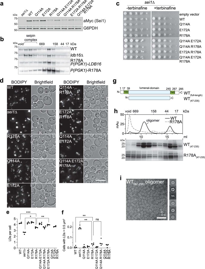
Extended Data Fig. 5. Lumenal domain interactions are mediated by R178.
(a) Western blot analysis with anti-myc antibodies of lysate from strains expressing WT seipin or indicated point mutations from the endogenous locus with C-terminal 13xmyc tag. (b) Size-exclusion chromatography of Triton X-100 solubilized membrane extracts of indicated strains expressing C-terminal 13xmyc-tagged seipin. Representative immunoblots of two biologically independent experiment repeats is shown. (c) Growth of yeast strain sei1∆ carrying vectors with C-terminally GFP-tagged SEI1 mutants or empty vector on synthetic medium ± 100 µg/ml terbinafine. (d) LD morphology of strains expressing indicated seipin mutants with C-terminal 13xmyc from endogenous locus. Size bar, 5 µm, (e,f) Quantification of experiment shown in d. n=4 biologically independent experiments. Data were analyzed with one-way ANOVA and Holm-Sidak’s posthoc comparisons; *, p<0.05; **, p<0.01; ***, p<0.001, ns, not significant. (g) Overview of lumenal domain construct purified from E. coli. (h) Size-exclusion chromatography analysis of affinity purified WT lumenal domain (WT(47-235)) or R178A(47-235). Top, traces of absorbance at 280 nm in mAu of WT and R178A lumenal domains. Bottom, SDS-PAGE analysis of 1-ml fractions by Coomassie staining. (i) Negative stain-EM analysis of WT lumenal domain oligomers shown in h. Right side shows 2D class averages. Size-bar, 500 Å.

