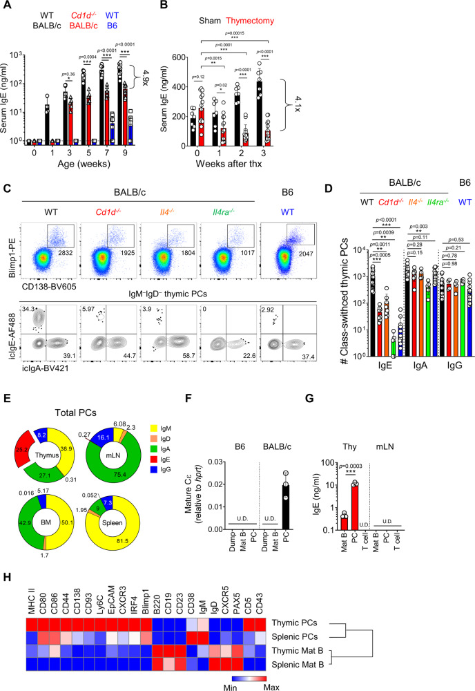
Fig. 1. IgE-producing PCs develop in the thymus.
A Graph shows serum IgE levels in B6 and WT and Cd1d−/− BALB/c mice measured by ELISA at the indicated ages (N = 6–9). Dotted lines indicate the detection limit. B Graph shows serum IgE concentrations in sham-operated and thymectomized BALB/c mice (N = 13) measured by ELISA. C, D Total thymocytes of B6 and BALB/c mice were enriched for CD138+ cells by MACS and stained with indicated markers. Representative dot plots are shown after gating out TCRβ+ cells (C). Numbers indicate the total number of cells in adjacent gates (upper) or the frequency of cells in each quadrant (lower). Graph shows numbers of icIgE, icIgA and icIgG+ PCs and statistical analysis (N = 4–19) (D). Results are from more than three independent experiments. E Pie charts show mean frequencies of each isotype of PCs in the thymus, mLN, BM, and spleen (N = 3–4). Numbers indicate mean values of frequencies. F, G Indicated cells were purified by MACS enrichment followed by FACS sorting. CD3+CD4+CD11b+ cells were used as dump cells. Graph shows relative levels of mature Cε transcripts normalized to hprt by qPCR (N = 3) (F). Indicated cells were cultured for 5 days, and total IgE concentrations were measured in the supernatant (N = 3) (G). H Heat map shows MFIs of indicated markers in B220+CD19+ mature B and PCs in thymus and spleen measured by flow cytometry. Data were log2 transformed and visualized by relative expression per column. Data are presented as mean values ± SD (A, B, D, F, and G). Each dot represents an individual mouse. Unpaired two-tailed t-test (A, B, and G) and one-way ANOVA (D) were used for comparison. ***p < 0.001, **p < 0.01, *p < 0.05. Not significant (p > 0.05). Thx Thymectomy; U.D. Undetected; PC Plasma cell; Mat B Mature B cell.

