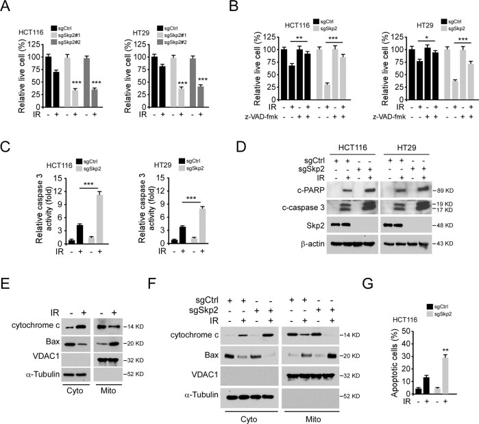
Fig. 2. Depletion of Skp2 enhances IR-induced intrinsic apoptosis.
A Skp2 knockout CRC stable cells were treated with/without IR (2 Gy) and cultured for 72 h, live cell population was determined by trypan blue exclusion assay. ***p < 0.001. B, C Skp2 knockout CRC stable cells were pretreated with pan-caspase inhibitor z-VAD-fmk for 4 h, followed by 2 Gy IR treatment. Cells were cultured for 72 h, live cell population was determined by trypan blue exclusion assay (B), caspase 3 activity was examined by Caspase 3 Assay Kit (C). *p < 0.05, **p < 0.01, ***p < 0.001. D Skp2 knockout HCT116 (left) and HT29 (right) stable cells were treated with/without IR (2 Gy) and cultured for 72 h, whole-cell extract (WCE) was subjected to IB analysis. E HCT116 cells were treated with/without IR (2 Gy) and cultured for 72 h, subcellular fractions were isolated and subjected to IB analysis. F, G Skp2 knockout HCT116 stable cells were treated with/without IR (2 Gy) and cultured for 72 h, subcellular fractions were isolated and subjected to IB analysis (F), the population of apoptotic cells was examined by flow cytometry (G). **p < 0.01.

