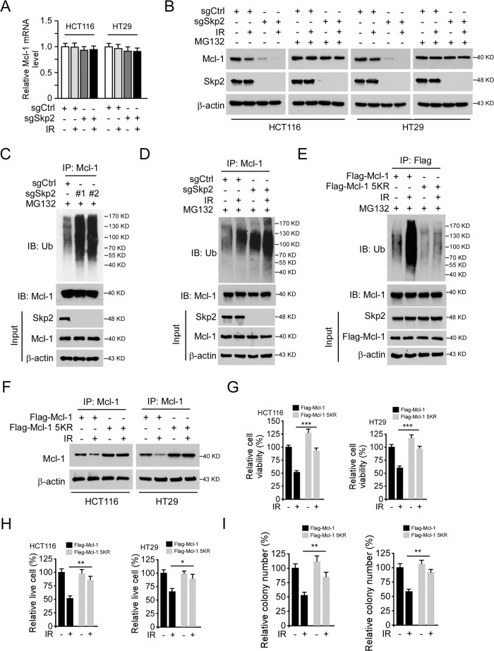
Fig. 4. Irradiation promotes Mcl-1 ubiquitination and degradation.
A Mcl-1 mRNA expression in Skp2 depleted CRC cells with IR treatment was examined by real-time RT-PCR. B Skp2 depleted HCT116, and HT29 cells were treated with IR (2 Gy) and cultured for 72 h, MG132 (25 μM) was added to the cell culture medium and maintained for 6 h. WCE was subjected to IB analysis. C Skp2 knockout HCT116 cells were treated with MG132 for 6 h, WCE was prepared and subjected to Mcl-1 ubiquitination analysis. D Skp2 depleted HCT116 cells were treated with MG132 (25 μM) for 6 h, followed by IR (2 Gy) treatment and cultured for 1 h. WCE was subjected to Mcl-1 ubiquitination analysis. E Flag-Mcl-1 wild type or 5KR mutant was transfected into HCT116 cells using lipofectamine 2000 for 48 h and treated with IR as indicated. WCE was extracted after IR treatment for 1 h and subjected to Mcl-1 ubiquitination analysis. F Flag-Mcl-1 wild type or 5KR mutant was transfected into HCT116 cells for 24 h, followed by IR (2 Gy) treatment, and cultured for 72 h. WCE was subjected to IB analysis. G–I Flag-Mcl-1 wild type or 5KR mutant was transfected into HCT116 and HT29 cells for 24 h, followed by IR (2 Gy) treatment and cultured for 72 h. Cell viability and live cell population were determined by MTS assay (G) and trypan blue exclusion assay (H), respectively. Colony formation was examined by plate colony formation assay (I). *p < 0.05, **p < 0.01, ***p < 0.001.

