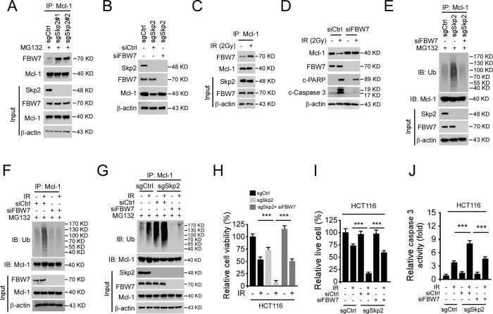
Fig. 5. FBW7 is required for IR-induced Mcl-1 ubiquitination.
A Skp2 knockout HCT116 cells were treated with MG132 for 6 h, WCE was subjected to co-immunoprecipitation (Co-IP) analysis. B siFBW7 was transfected into Skp2 depleted HCT116 cells and subjected to IB analysis. C HCT116 cells were treated with IR (2 Gy), WCE was collected 1 h later and subjected to Co-IP analysis. D siFBW7 was transfected into HCT116 cells for 24 h, followed by IR (2 Gy) treatment. Cells were cultured for 72 h, WCE was subjected to IB analysis. E siFBW7 was transfected into Skp2 depleted HCT116 cells for 48 h, followed by MG132 treatment for 6 h, WCE was subjected to IP-mediated Mcl-1 ubiquitination analysis. F HCT116 cells were transfected with siCtrl or siFBW7 and cultured for 48 h. After being incubated with MG132 for 6 h, the cells were treated with IR. WCE was collected 1 h later and subjected to IP-mediated Mcl-1 ubiquitination analysis. G–J Skp2 depleted HCT116 cells were transfected with siCtrl or siFBW7 and cultured for 48 h. After being incubated with MG132 for 6 h, the cells were treated with IR. WCE was collected 1 h later and subjected to IP-mediated Mcl-1 ubiquitination analysis (G). Cell viability and live cell population was determined by MTS assay (H) and trypan blue exclusion assay (I), respectively. Caspase 3 activity was examined by Caspase 3 Assay Kit (J). ***p < 0.001.

