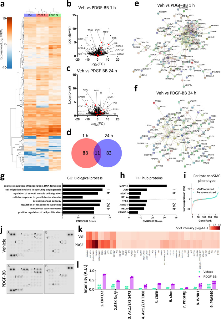
Fig. 2. PDGF-BB activates a biphasic response in brain pericytes.
Pericytes derived from neurologically normal post-mortem patients were treated with vehicle or PDGF-BB (10 ng/mL) for 1 or 24 h and gene expression analysed by RNAseq. a Dendrogram of PDGF-BB response in pericytes. Volcano plots comparing b 1 h and c 24 h PDGF-BB treatment to vehicle, with hits highlighted. d Venn diagram of differentially expressed gene lists at 1 and 24 h of PDGF-BB treatment. STRING network analysis of hits from e 1 and f 24 h of PDGF-BB treatment. g Gene ontology analysis and h interaction hub analysis of differentially expressed gene lists. i Expression of pericyte and vascular smooth muscle cell lineage markers identified by Vanlandewijck et al.15, ranked by fold change. Pericytes derived from the middle temporal gyrus of epilepsy patients were serum starved overnight, then treated with either PDGF-BB (100 ng/mL) or vehicle and lysed. Phosphorylation was detected in lysates using an antibody array. j Representative images, k heatmap of all phosphorylation sites and l levels of selected phosphorylation events in vehicle or PDGF-BB-treated pericytes.

