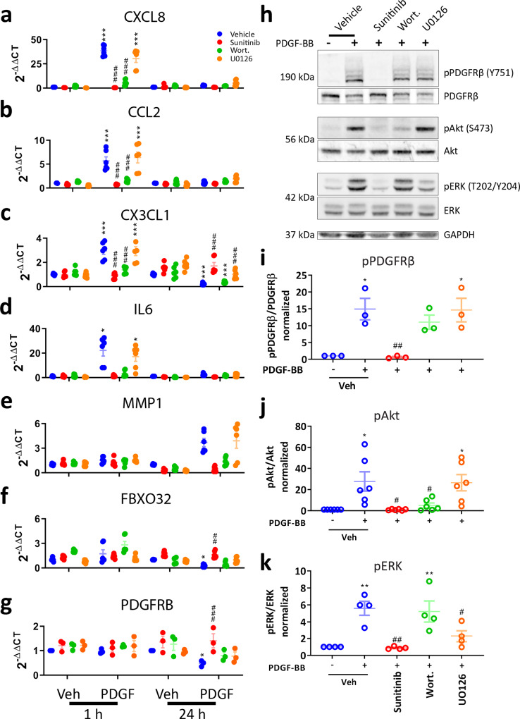
Fig. 5. Akt and ERK activate distinct aspects of the PDGF-BB response in pericytes.
Pericytes were incubated with vehicle (0.3% DMSO), PDGFRβ inhibitor sunitinib (100 nM), PI3K inhibitor wortmannin (100 nM) or MEK/ERK inhibitor U0126 (10 μM) for 30 min. Pericytes were then treated with either vehicle or PDGF-BB (10 ng/mL) for 1 or 24 h, and RNA extracted for qPCR. Expression of a CXCL8, b CCL2, c CX3CL1, d IL6, e MMP1, f FBXO32 and g PDGFRB in pericytes treated with PDGF-BB for 1 or 24 h, with or without pathway inhibitors. n = 6, two-way ANOVA. Pericytes were serum starved overnight, then pre-treated with sunitinib (100 nM), wortmannin (100 nM) or U0126 (10 μM) or vehicle (0.3% DMSO) for 30 min, then treated with vehicle or PDGF-BB (100 ng/mL) for 30 min. Cells were lysed and western blot was performed. h Representative blots and densitometric analysis of i PDGFRβ, j Akt and k ERK phosphorylation. n = 4, one-way ANOVA. *p < 0.05, **p < 0.01, ***p < 0.001 vs vehicle control, #p < 0.05, ##p < 0.01, ###p < 0.001 vs PDGF-BB-treated.

