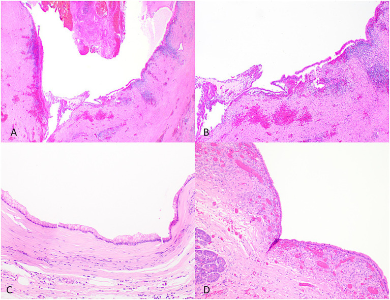
Figure 4.
Representative histological pictures of retention cyst, simple mucinous cyst, and mucinous cystic neoplasm. (A) Retention cyst. Low magnification shows a dilated pancreatic duct. (B) Higher magnification shows a flattened epithelium with no papillary projections. Note the background pancreas with extensive atrophy and fibrosis. No ovarian-type stroma is seen. (C) Simple mucinous cyst. The cyst is lined by a benign mucinous epithelium and lacks an ovarian stroma. (D) Mucinous cystic neoplasm. The cyst is lined by mucinous epithelium and unlike simple mucinous cyst or a retention cyst is associated with an ovarian-type stroma. (A) Original magnification 10×; (B) Original magnification 100×; and (C,D) Original magnification 200×.

