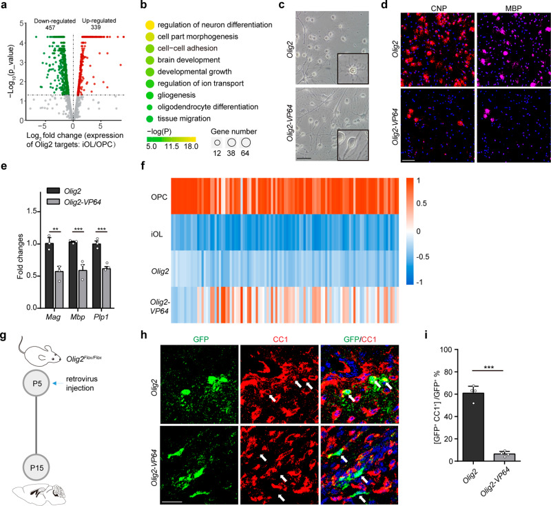
Fig. 1. The repressive role of OLIG2 is essential for oligodendrocyte differentiation.
a Volcano plot of differential expressed genes (DEGs, log2FC ≥ 1 or ≤ -1; p value <0.05) that are targeted by enhanced OLIG2 binding in rat OPCs to iOL transition. b Representative GO analysis of the significantly downregulated genes in Fig. 1a. c, d Representative phase micrographs(c) and CNP+ and MBP+ immunostaining (d) in rat OPCs transfected with Olig2 or Olig2-VP64 under differentiation condition. n = 3 independent experiments. e Quantitative real-time PCR analysis of oligodendrocyte differentiation-associated genes in rat OPCs transfected with Olig2 or Olig2-VP64 under differentiation condition. n = 3 independent experiments. Error bars indicate SEM (Mag, p = 0.0084; Mbp, p < 0.001; Plp, p < 0.001. two-tailed unpaired Student’s t test). f Heat map representing the expression of OL differentiation–related genes in rat OPC, iOL, iOL transfected with OLIG2 and iOL transfected with OLIG2-VP64. g Schematic diagram for retrovirus injection. h Immunostaining for CC1 on OLIG2 or OLIG2-VP64 retrovirus infected brain at P15. Arrow indicates the cell infected with retrovirus. n = 4 control and 4 Olig2-VP64 mice. i Quantification of the percentage of differentiated oligodendrocytes in infected cells. Error bars indicate SEM (p < 0.001. two-tailed unpaired Student’s t test). Scale bars: 50 μm in (c); 100 μm in (d); 25 μm in (h).

