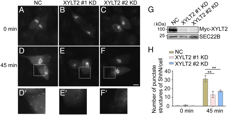
Fig. 4.
Synthesis of PGs regulates export of ShhN out of the TGN. (A–F) HeLa cells were transfected with NC siRNA or two different siRNAs against XYLT2. At 48 h after transfection, cells were retransfected with plasmids encoding Str-KDEL and SBP-EGFP-ShhN25-198. On day 3 after knockdown, cells were treated with biotin and incubated in the 20 °C for 2 h. Then the cells were incubated at 32 °C for 0 or 45 min, and the localization of Shh was analyzed (Scale bar, 10 μm). Magnification, 63× . The magnified views of the indicated area in panels D–F are shown in panels D′–F′. (G) HEK293T were transfected with negative control (NC) siRNA or siRNA against XYLT2. At 48 h after transfection, cells were retransfected with plasmids encoding Myc-XYLT2. On day 3 after knockdown, the level of SEC22B and Myc-XYLT2 in cell lysates was analyzed by immunoblotting using anti-Myc or anti-SEC22B antibodies. (H) Quantifications of the number of punctate structures containing SBP-EGFP-ShhN25-198 per cell at different time points after biotin treatment (n = 3, mean ± SD, over 20 cells were quantified in each experimental group). **P < 0.01.

