Fig. 1.
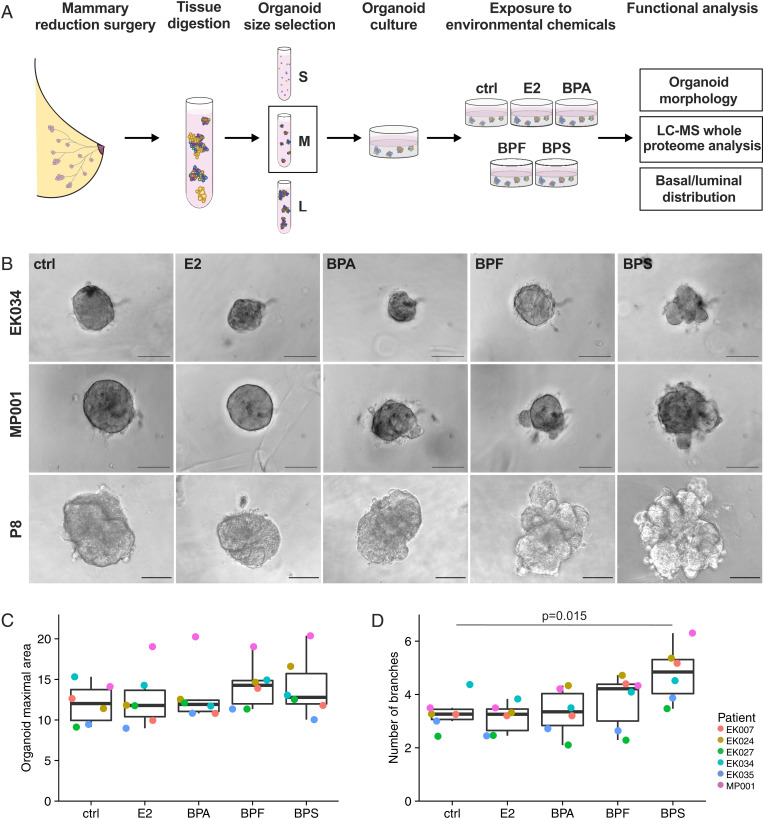
Bisphenol exposures alter branching morphology of human mammary organoids. (A) Schematic of the experimental workflow. Nonmalignant primary human mammary tissue from reduction mammoplasty surgeries was digested and size separated, and the medium (M)-sized (vs. small [S] or large [L]) organoids were cultured in Matrigel. They were exposed to the vehicle control (ctrl), E2, or one of the bisphenols (15 nM) for 6 d. The end points analyzed included organoid morphology, the proteome at a global level (liquid chromatography-mass spectrometry [LC-MS]), and the distribution of basal and luminal cells. (B) Representative bright-field images of human breast organoids of different patients after exposure for 6 d to the vehicle control (ctrl), E2, BPA, BPF, or BPS. (Scale bars, 100 μm.) (C) Quantification of the organoid maximal cross-sectional area colored by patient (cohort A). Each dot represents the mean value from the analysis of 5 to 25 organoids per patient per treatment. The median and the interquartile range are shown. The values were not statistically different among the groups. (D) Quantification of the total number of branches per organoid colored by patient (cohort A). Each dot represents the mean number of branches from 5 to 25 organoids per patient per treatment. The median and interquartile range are shown. Results that reached statistical significance using the two-sample Wilcoxon test are noted.

