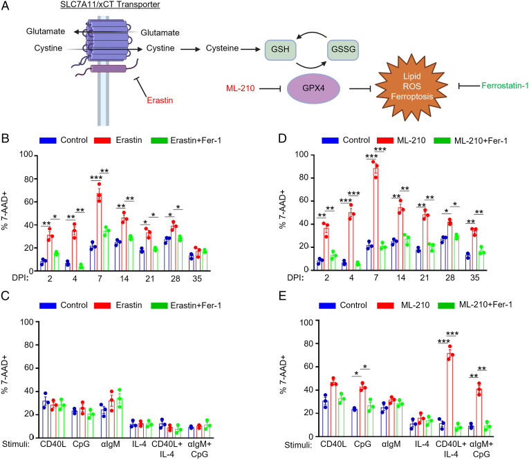
Fig. 2.
Effects of EBV infection versus B cell receptor stimulation on sensitization to ferroptosis. (A) Model of erastin and ML-210 ferroptosis inducing agent mechanisms of action on ferroptosis induction. GSH, reduced glutathione. GSSG, oxidized glutathione. (B) FACS %7-AAD+ mean + SD values from n = 3 independent replicates of primary B cells from independent donors infected by EBV for the indicated day postinfection (DPI) and treated with 10 μM erastin and/or 5 μM Fer-1 for 24 h prior to analysis as indicated. (C) FACS %7-AAD+ mean + SD values from n = 3 independent replicates of primary B cells from independent donors stimulated as indicated by Mega-CD40L (50 ng/mL), αIgM (1 μg/mL), CpG (1 μM), or IL-4 (20 ng/mL) for 48 h and treated with 10 μM erastin and/or 5 μM Fer-1 for 24 h prior to analysis as indicated. (D) FACS %7-AAD+ mean + SD values from n = 3 independent replicates using primary B cells from independent donors infected by EBV for the indicated DPI and treated with 1 μM ML-210 and/or 5 μM Fer-1 for 24 h prior to analysis as indicated. (E) FACS %7-AAD+ mean + SD values from n = 3 independent replicates of primary B cells from independent donors stimulated as indicated for 48 h and treated with 1 μM ML-210 and/or 5 μM Fer-1 for 24 h prior to analysis as indicated. P values were determined by one-sided Fisher’s exact test. *P < 0.05, **P < 0.005, ***P < 0.0005.

