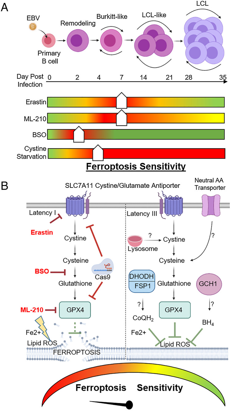
Fig. 7.
Model of EBV effects on B cell vulnerability to ferroptosis. (A) Schematic of vulnerability to ferroptosis induction at distinct states of EBV-mediated B cell transformation. Ferroptometer sliders demarcate the relative sensitivity of EBV-infected cells to ferroptosis induction to the indicated agent, with green and red indicating the lowest and highest sensitivities to ferroptosis induction, respectively. (B) Schematic of vulnerability to ferroptosis-inducing agents in latency I (Left) versus III (Right) EBV-transformed B cells. Question marks indicate potential pathways by which latency III cells may differentially acquire cysteine and buffer lipid ROS for redox defense.

