Figure 1.
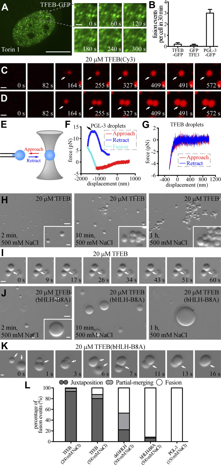
TFEB forms phase-separated condensates with low fusion propensity. (A and B) Time-lapse analysis showing that the nuclear TFEB-GFP puncta in Torin 1–treated HeLa cells rarely fuse with each other (A). Quantification of fusion events in each cell during a 30 min period for TFEB-GFP, GFP-TFE3, and PGL-3-GFP droplets (B). Data are shown as mean ± SEM (n = 10 cells for each bar) in B. (C and D) Cy3-labeled TFEB droplets grow with time (white arrow) in a buffer containing 200 mM NaCl (C). Upon encounter (white arrow), two TFEB droplets fail to fuse during the period of examination (D). The time refers to imaging time, and the time point of the first image is defined as 0 s. (E) Schematic for the optical tweezers experiments. A single droplet is held on a micropipette by suction, and another droplet is trapped by the optical tweezers. The force between the two droplets during multiple approach and retraction phases is measured by the optical tweezers and recorded. Droplets with a diameter of ∼5–10 μm were used for the experiments. (F and G) Representative force–displacement curves recording the force between two PGL-3 droplets formed in 150 mM NaCl buffer (F) or TFEB droplets formed in 500 mM NaCl buffer (G) during approach and retraction. Repulsive forces are detected when two PGL-3 droplets get close. The two PGL-3 droplets fuse together at approximately −1,000 nm, and the repulsive forces gradually convert to attractive forces due to shrinkage of the fused droplets. At the top of the blue curve (arrow), the optical tweezers fell off the trapped droplet (F). No obvious forces are detected before contact between two TFEB droplets (G). The indentations on TFEB droplets are much smaller than on PGL-3 droplets at the force of approximately −4 pN, which suggests that TFEB droplets are much harder than PGL-3 droplets. (H and I) DIC images showing that 20 μM TFEB forms droplets in 500 mM NaCl buffer 2 min after LLPS induction. The size of droplets remains unchanged from 10 min to 1 h after induction. The droplets gradually cluster with time (H). Time-lapse images of two encountering TFEB droplets that do not undergo fusion with time (I; arrows). The time point of the first image is defined as 0 s. (J and K) DIC images showing that 20 μM TFEB(bHLH-B8A) forms droplets in 500 mM NaCl buffer from 2 min to 1 h after LLPS induction (J). The droplets continuously fuse upon encounter, resulting in formation of large droplets (J). Time-lapse images of three encountering TFEB(bHLH-B8A) droplets which undergo rapid fusion (K; arrows). The time point of the first image is defined as 0 s. (L) Percentage of fusion events for droplets formed by 20 μM wild-type and mutant TFEB and 10 μM PGL-3 in the indicated salt buffers. Three types of event (juxtaposition, partial merging, and fusion) are defined for droplets that underwent an encounter for 30 s. 124, 106, 113, 106, and 100 fusion events were analyzed for TFEB in 200 and 500 mM NaCl buffer; TFEB(del bHLH) and TFEB(bHLH-B8A) in 500 mM NaCl; and PGL-3 in 150 mM NaCl, respectively. Scale bars: 10 μm (A); 5 μm (H and J); 1 μm (C, D, I, and K); 1 μm (enlarged figures in A and insets in H and J).

