Figure 2.
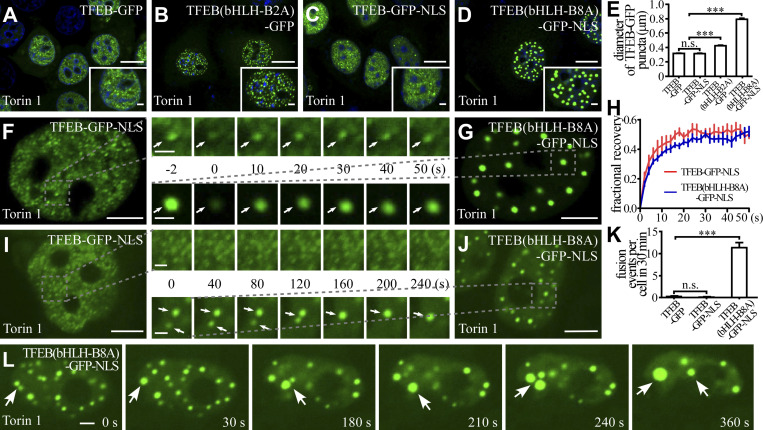
The effect of charged residues in the bHLH domain on the formation and biophysical properties of TFEB puncta in living cells. (A–E) Puncta formed by expressing wild-type or mutant TFEB-GFP and TFEB-GFP-NLS upon Torin 1 treatment in the nucleus of HeLa cells. Compared with TFEB-GFP puncta (A), the TFEB(bHLH-B2A)-GFP puncta are larger (B). The size of TFEB-GFP-NLS puncta is similar to TFEB-GFP puncta (C), while the size of TFEB(bHLH-B8A)-GFP-NLS droplets is much larger (D). Quantification of the diameter of nuclear wild-type or mutant TFEB-GFP and TFEB-GFP-NLS puncta (E). The biggest 10 puncta in each cell were chosen, and the diameters were measured by ImageJ. Data are shown as mean ± SEM (n = 100 puncta for each bar). ***, P < 0.001. (F–H) FRAP analysis of the TFEB-GFP-NLS (F) and TFEB(bHLH-B8A)-GFP-NLS (G) signals of the punctate structures (arrows) in the nucleus of Torin 1–treated HeLa cells. Quantification of the FRAP data for F and G (H). Data are shown as mean ± SEM (n = 3) in H. (I–K) Time-lapse experiments showing that TFEB-GFP-NLS puncta rarely fuse with each other (I), while TFEB(bHLH-B8A)-GFP-NLS puncta (arrows) undergo frequent fusion (J) in the nucleus of Torin 1–treated HeLa cells. Quantification of fusion events in each cell for 30 min for TFEB-GFP, TFEB-GFP-NLS, and TFEB(bHLH-B8A)-GFP-NLS droplets (K). Data are shown as mean ± SEM (n = 10 cells for each bar) in K. ***, P < 0.001. (L) Nuclear TFEB(bHLH-B8A)-GFP-NLS puncta in Torin 1–treated HeLa cells undergo continuous fusion (arrows) and become larger with time. Scale bars: 10 μm (A–D); 5 μm (F, G, I, and J); 2 μm (L and enlarged figures in F and G); 1 μm (enlarged figures in I and J and insets in A–D).

