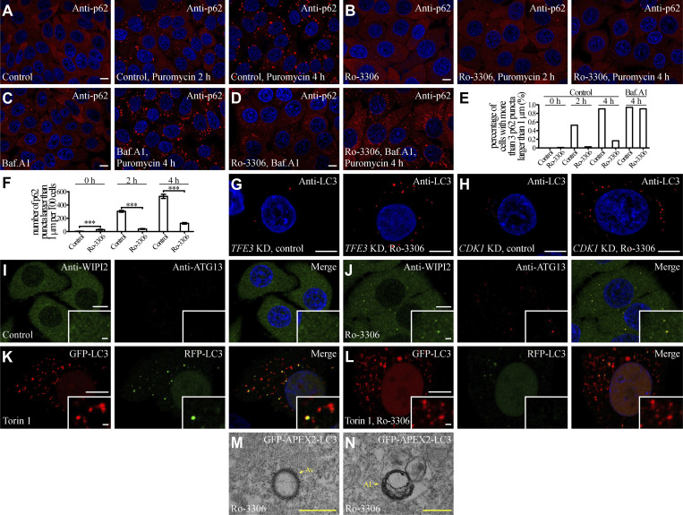
Figure S5.
Ro-3306 treatment promotes autophagy. (A–F) In puromycin-treated HeLa cells, p62 (detected by anti-p62 antibody) forms an increasing number of aggregates with time (A). The number of p62 aggregates is dramatically reduced by treatment with 10 μM Ro-3306 (B), and is restored by simultaneous incubation with Baf.A1 (C and D). (E and F) Quantification of the percentage of cells with more than three p62 puncta larger than 1 μm (n > 200 cells from at least three independent experiments; E), and the number of p62 puncta larger than 1 μm per 100 cells (F) in control and Ro-3306 treated cells in response to puromycin treatment with the indicated time. Data are shown as mean ± SEM in F (n > 200 cells from at least three independent experiments in F). ***, P < 0.001. (G and H) The number of LC3 puncta stained by anti-LC3 is significantly increased by 10 μM Ro-3306 treatment in TFE3 KD (G) and CDK1 KD (H) HeLa cells. (I and J) Compared to control cells (I), the number of endogenous WIPI2 and ATG13 puncta stained by anti-WIPI2 and anti-ATG13 antibodies is higher in 10 μM Ro-3306-treated HeLa cells (J) under normal conditions. (K and L) The RFP-GFP-LC3 assay in control and 10 μM Ro-3306-treated HeLa cells after Torin 1 treatment. Compared to control cells (K), more red-only puncta are formed in Ro-3306-treated cells (L). (M and N) TEM images showing that dark APEX2-LC3-derived signals are detected on the membranes of autophagosomes (M) and autolysosomes (N) in 10 μM Ro-3306-treated HeLa cells. AL, autolysosome; Av, autophagosome. Scale bars, A–D and G–L, 10 μm; inserts in I–L, 1 μm; M and N, 500 nm.

