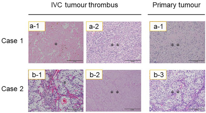
Figure 2. Microscopic appearance of inferior vena cava (IVC) tumour thrombi and primary tumours after combination treatment with immune checkpoint inhibitors. The two haematoxylin and eosin-stained sections on the left were taken from IVC tumour thrombi and those on the right are from primary tumours. Residual malignant cells are present (double asterisk) in these tumours, as was the necrotic tissue that replaced the IVC tumour thrombus (asterisk). Scale bar=2 mm.

