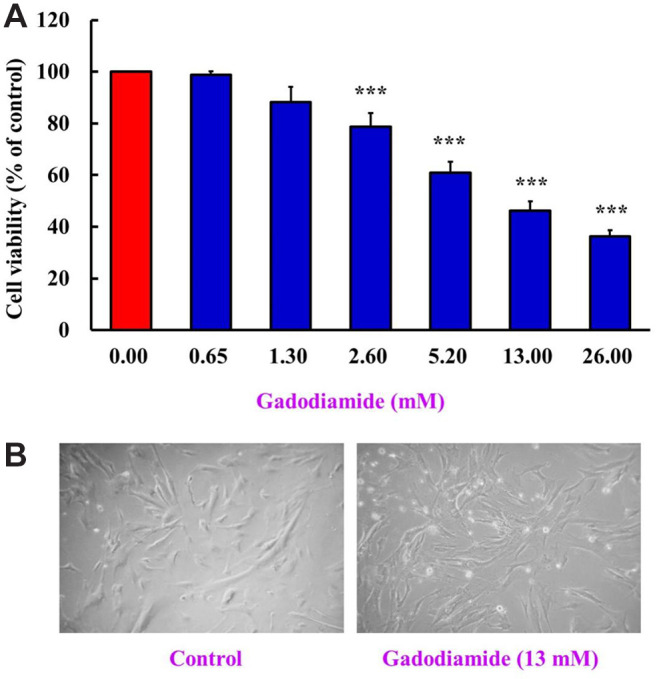
Figure 1. Gadodiamide suppressed HaCaT cell viability. (A) HaCaT cells (5×10⁴ cells/well) were incubated for 24 h with 0, 0.65, 1.3, 2.6, 5.2, 13, or 26 mM of gadodiamide. HaCaT cell viability was assessed using the MTT assay. The results are normalized using the vehicletreated control as 100% (n=3; ***p<0.05). (B) Cell morphology of HaCaT cells was observed after treatment with 13 mM gadodiamide for 24 h. Left: vehicle-treated control; right: 13 mM gadodiamidetreated cells.