. 2022 Mar 3;36(2):582–595. doi: 10.21873/invivo.12741

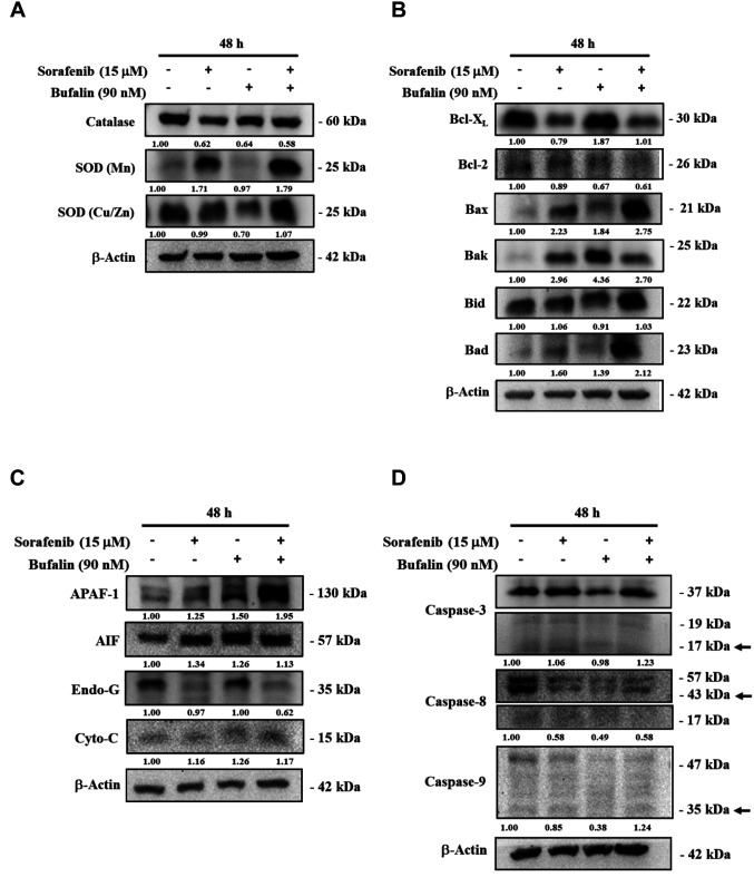
Figure 5. The effects of sorafenib, bufalin, or the combination of sorafenib with bufalin on the ROS- or apoptosis-associated proteins in NCI-H292 cells. Cells (1×10⁶ cells/well) were subjected to 15 μM sorafenib, 90 nM bufalin, or the combination of 15 μM sorafenib with 90 nM bufalin for 48 h. Cells were collected and lysed, and their total proteins were quantified and analyzed by western blotting. The blotting was probed with primary antibodies against anti-SOD (Mn), -SOD (Cu/Zn), and -catalase (A); -Bcl-XL, -Bcl-2, -Bax, -Bak, -Bid, and –Bad (B); -APAF-1, -AIF, -Endo G, and -Cytochrome C (C); -caspase-3, -caspase-8, and -caspase-8 (D). Then the membranes were washed, followed by incubation with responding secondary antibody (anti-mouse or anti-rabbit antibodies). The protein signals were detected by ECL and quantified by the ImageJ software.

Figure 5