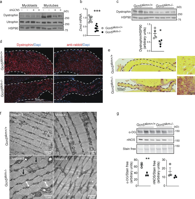
Figure 2.
GCN5 regulates dystrophin protein expression and muscle integrity. (a) Western blot of protein extract from C2C12 myoblasts and myotubes (n = 2/condition) after 4 d of differentiation in the presence or absence of adenovirus-driven shGCN5 using anti-dystrophin and anti-utrophin antibodies, with anti-HSP90 antibodies used as a loading control. (b) Relative expression of Dmd in gastrocnemius muscle from 6-mo-old control and Gcn5skm−/− mice as measured by RT-qPCR relative to housekeeping genes 36b4 and Gapdh. Data are presented as mean ± SEM. n = 8–11/group. ***, P < 0.01 versus Gcn5skm+/+ as measured by two-tailed Student’s t test. (c) Western blot of whole protein extract from tibialis anterior muscle of 6-mo-old control and Gcn5skm−/− mice using anti-dystrophin with anti-HSP90 antibodies used as a loading control. Data are presented as mean ± SEM. n = 4/group. *, P < 0.05 versus Gcn5skm+/+ as measured by two-tailed Student’s t test. (d) Representative immunofluorescence images of diaphragm muscle sections from 6-mo-old control and Gcn5skm−/− mice using anti-dystrophin (left column) or nonspecific anti-rabbit secondary (right column) antibodies. Images are tile scanned and stitched. Dashed line represents the edge of liver tissue, which was used to mount the diaphragm tissues for sectioning. n = 3/group. Scale bar, 500 μm. (e) Picrosirius red staining of diaphragm muscle sections from 6-mo-old control and Gcn5skm−/− mice. Images were tile scanned and stitched. Dashed line represents the edge of liver tissue that was used on both sides of the diaphragm to help with tissue mounting and sectioning. n = 3/group. Scale bars, 500 μm; inset, 20 μm. (f) 8-wk-old Gcn5skm−/− mice harbor variable abnormal sarcomere structures, including loss of rectangular sarcomere structure (white double-ended arrows), offset z-discs (black arrows), and/or disrupted sarcomeres (white asterisks) as revealed on electron transmission micrographs of EDL muscles. No steps were taken to prevent muscle contraction during sacrifice. (g) Western blot of whole protein extract from left quadriceps muscles of 6-mo-old control and Gcn5skm−/− mice using anti–α-dystroglycan (α-DG) and anti-nNOS antibodies and stain-free protein loading control. Data are presented as mean ± SEM. n = 3/group. **, P < 0.01 versus Gcn5skm+/+ as measured by two-tailed Student’s t test.

