
Fig. 6, Movie 8. Vesicle-like structures containing procollagen and HSP47 inside ER lumen and lysosomal membranes.
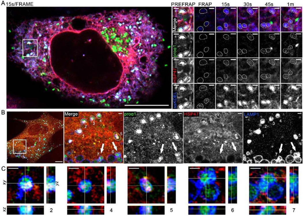
(A, Movie 8) Airyscan single slice frames and time-lapse video of colocalization of Venus-proα1(I), Cherry-HSP47, and ssCFP-KDEL in vesicle-like structures inside ER lumen and fluorescence recovery of these markers after photobleaching (FRAP); N=18 (3 experiments). Zoomed still frames show the same area as the inset in Movie 8. In these zoomed frames, 3 vesicle-like structures marked by Venus-proα1(I), Cherry-HSP47, and ssCFP-KDEL are traced by thin white lines. One of these structures is also highlighted by a white circle in the movie inset. The bright green structure in 45 s and 1 min frames is likely a procollagen transport vesicle containing no Cherry-HSP47 and no ssCFP-KDEL, which entered the field of view between 30 and 45 s post-bleaching (the 15s/frame imaging rate in this experiment was not fast enough for more definitive identification). Quantitative analysis of FRAP kinetics in shown in Fig. 7C with and without BFA treatment. (B, C) 3D Airyscan imaging of Venus-proα1(I) and Cherry-HSP47 colocalization in lysosomes/late endosomes marked by membrane protein LAMP1-CFP; N=7. (B) A slice from the 3D z-stack shows whole cell and zoomed images of vesicle-like structures 1–7, in which Venus-proα1(I) is colocalized with LAMP1-CFP. (C) Orthogonal cross-sections of structures 2, 4, 5, 6, and 7. Structures 2, 4, and 5 were likely formed by direct lysosomal engulfment of ERES [37] or ER lumen [44], since they are surrounded by LAMP1-positive lysosomal membrane and contain internalized procollagen, HSP47, and LAMP1. Lysosomes 6 and 7 contain little or no internalized HSP47 and thereby might have a different origin, e.g. endocytosis of secreted procollagen molecules. Scale bars = 10 μm (whole cell) and = 1 μm (zoom).