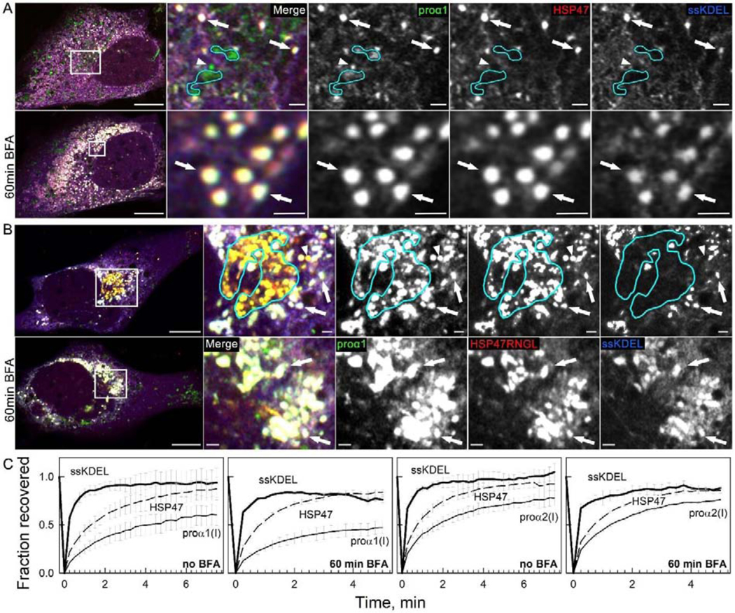
Fig. 7, Movie 9. Golgi disruption by brefeldin A (BFA) causes accumulation of vesicle-like regions of ER lumen filled with procollagen and HSP47, which were previously misinterpreted as procollagen transport vesicles.

(A) Colocalization of Venus-proα1(I), Cherry-HSP47, and ssCFP-KDEL in vesicle-like structures inside ER lumen (white puncta in merged images marked by arrows) and accumulation of these structures after 60 min treatment of the same cell with 5 μg/ml BFA (bottom panels); N=9. Golgi cisternae (outlined by cyan lines) and likely transport vesicle (arrowhead) contain Venus-proα1(I) but not Cherry-HSP47 or ssCFP-KDEL. They disappear rather than accumulate after BFA treatment. (Movie 9) Time-lapse video of similar Venus-proα1(I)/Cherry-HSP47/ssCFP-KDEL puncta in a different cell after BFA treatment shows nearly instantaneous ssCFP-KDEL fluorescence recovery after photobleaching, demonstrating that these puncta are integrated within the ER lumen network. (B) Similar accumulation of Venus-proα1(I), Cherry-HSP47RNGL, and ssCFP-KDEL in vesicle-like white puncta (arrows) inside ER lumen after 60 min treatment with 5 μg/ml BFA; N=8. Golgi cisternae (outlined in cyan) and likely transport vesicle (arrowhead) contain Venus-proα1(I) and Cherry-HSP47RNGL but not ssCFP-KDEL. They disappear after BFA treatment. (C) FRAP kinetics in vesicle-like structures containing colocalized ssCFP-KDEL, Venus-proα1(I)/proα2(I), and Cherry-HSP47; N=3. The images in (A,B) are Airyscan single slices; scale bars = 10 μm (whole cell) and = 1 μm (zoom).
