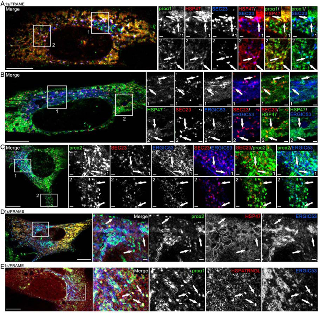
Fig. 8, Movies 10–12. HSP47 entry into ERES and release at ERES regions marked by ERGIC53.

(A, Movie 10) Airyscan single slice frame and time-lapse video of GFP-proα1(I), Cherry-HSP47, and TagBFP2-SEC23 colocalization in punctate structures representing ERES (arrows) in the Golgi region (Movie inset and zoomed still frame in top panels) and away from the Golgi region (zoomed still frame in bottom panels). Based on larger size and slower motion, procollagen carrier appearing at 00:28 s frame in the movie inset is more likely a secretory vesicle than ER-Golgi transport intermediate. N=20 (3 experiments). (B) Airyscan slice of similar colocalization of Venus-HSP47, Halo-SEC23 and Cerulean-ERGIC53 at ERES (arrows); N=6. (C) Airyscan slice of similar colocalization of Venus-proα2(I), Halo-SEC23, and Cerulean-ERGIC53 at ERES (arrows); N=22 (5 experiments). (D, Movie 11) Airyscan single slice frame and time-lapse video of Venus-proα2(I) and Cerulean-ERGIC53 colocalization without Cherry-HSP47 in procollagen transport vesicles (arrows) and Golgi (white outlines); N=4. (E, Movie 12) Airyscan single slice frame and time-lapse video of Venus-proα1(I), Cherry-HSP47RNGL, and Cerulean-ERGIC53 colocalization in procollagen transport vesicles (arrows) and Golgi (white outlines); N=17 (3 experiments). Scale bars = 10 μm (whole cell) and = 1 μm (zoom).
