Fig. 6.
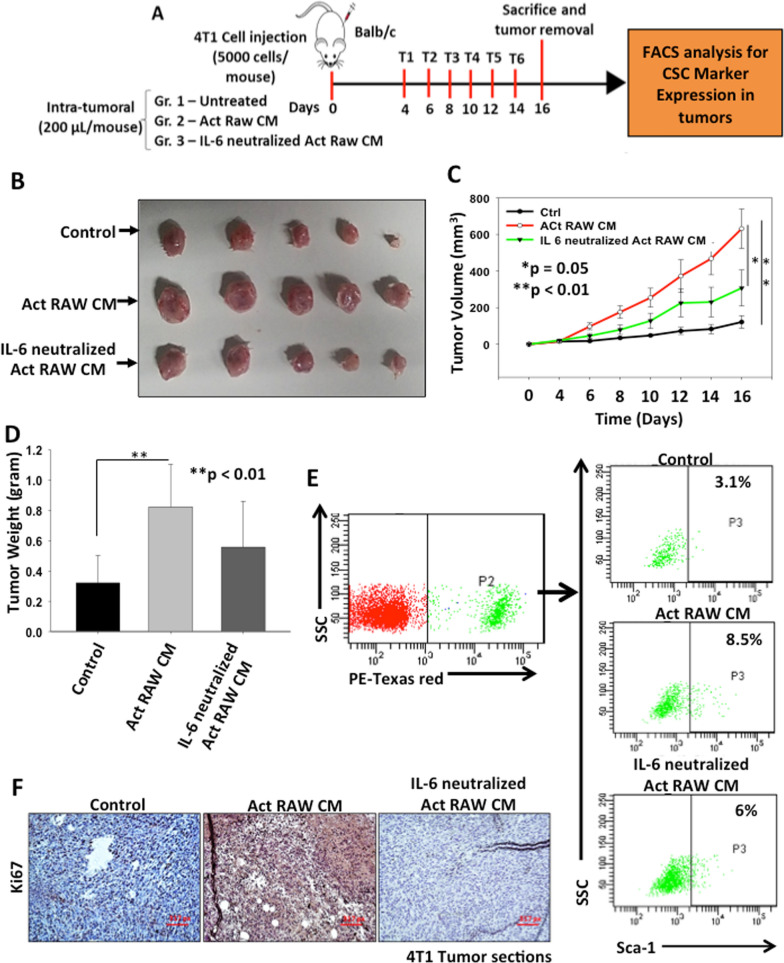
TAMs derived IL-6 promotes tumor growth in breast cancer. A Schematic representation of experimental plan. Briefly, 4T1 cells were injected into the mammary fat pad of female BALB/c mice and when the tumor reached palpability, mice were treated with either CM of activated RAW264.7 cells or IL-6 neutralized CM of activated RAW264.7 cells and tumor growth was observed. After treatments, mice were sacrificed, tumors were excised and tumor weight and volume were observed. B Digital photographs of excised tumors. C Tumor volumes were calculated and analysed statistically. The line graph describes change in mean tumor volume with respect to time in different groups. Data is represented in mean ± SE, *denotes p < 0.05, **denotes p < 0.01, n = 5. D Tumors were excised, weighed and analysed statistically. Bar graph represents mean tumor weight ± SD, **denotes p < 0.01, n = 5. E FACS analysis was performed to check the CSC population from these tumors. Breast cancer cells population was selected based on their endogenous expression of tdTomato fluorescent protein and expression of Sca-1 was examined in these populations using FACS analysis. F Expression of Ki67 in control or Act RAW CM and IL-6 neutralized Act RAW CM treated 4T1 tumor sections examined by immunohistochemistry

