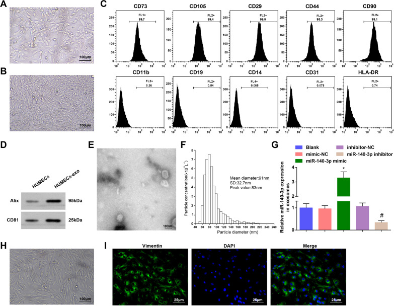
Fig. 1.
Identification of HUCMSCs, HUCMSCs-exo and RASFs. A Morphological observation of low density HUMSCs; B Morphological observation of high density HUMSCs ; C Surface antigens of HUMSCs were detected by flow cytometry; D Expression of CD81 and Alix in exosomes was determined by Western blot analysis; E Exosomes were identified through a TEM; F Particle size of exosomes was analyzed by NTA; G Expression of exosomal miR-140-3p was assessed by RT-qPCR; H Morphological observation of rat RASFs ; I Rat RASFs were identified by vimentin protein immunofluorescent staining; N = 3; *P < 0.05 vs the mimic-NC group, #P < 0.05 vs the inhibitor-NC group; the data were expressed as mean ± standard deviation, ANOVA was used for comparisons among multiple groups and Tukey’s post hoc test was used for pairwise comparisons after ANOVA

