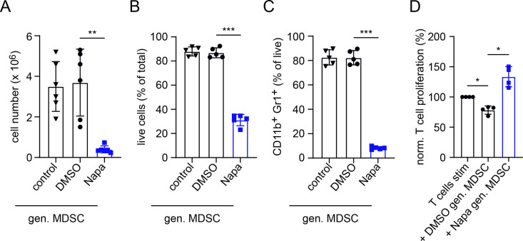
Figure 4.
Inhibition of MDSC generation in vitro by Napaucasin. MDSCs were generated in vitro with IL-6 and GM-CSF (40 ng/mL each, control). Napa (1 µM) or DMSO (0.01%) was added together with cytokines. (A) Data are shown as cell numbers per plate after 4 days of incubation (n=6). Results are presented as the percentage of live (7AAD–) cells within total cells (B) and the percentage of CD11b+Gr1+ cells among total cells (C) measured by flow cytometry (mean±SD, n=5). (D) The function of in vitro generated MDSC was determined on the coculture with CFSE-labeled murine-activated splenic CD8+ T cells. After 72 hours of incubation, T-cell proliferation was evaluated by CFSE dilution measured by flow cytometry. Cumulative data for T-cell proliferation are presented as the percentage of divided T cells norm. to the respective control of stimulated T cells alone (mean±SD, n=4). Statistics were performed on not norm. data. *P<0.05,**P<0.01, ***P<0.001. CFSE, carboxyfluorescein succinimidyl ester; gen., generated; GM-CSF, granulocyte–macrophage colony-stimulating factor; IL, interleukin; MDSC, myeloid-derived suppressor cell; Napa, Napabucasin; norm., normalized.

