
Figure 2.
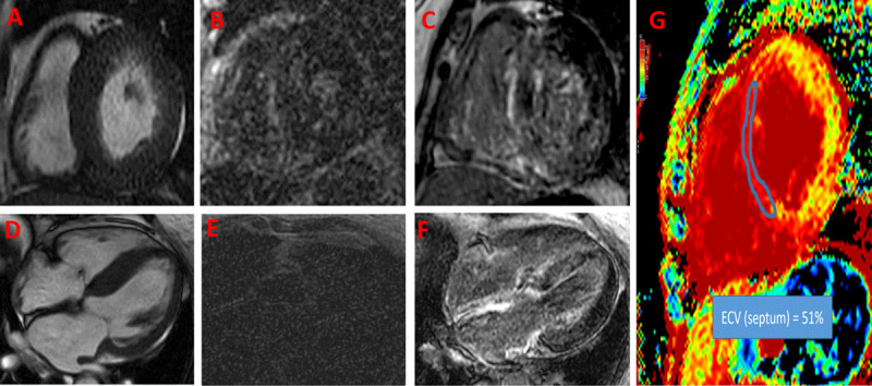
Cardiac magnetic resonance imaging (CMR) of a patient presenting with dyspnea and found to have left ventricular hypertrophy. First set of images (A, B and E) were obtained prior to presenting to our practice. Image A shows concentric left ventricular hypertrophy. Due to abnormal gadolinium kinetics and selecting an inappropriately low inversion time, late gadolinium enhancement (B) short axis and (E) 4-chamber views were not interpretable. (D) Repeat CMR shows severe asymmetrical septal hypertrophy on 4-chamber view; with choosing an appropriate inversion time for late gadolinium enhancement imaging, there was global enhancement of the left ventricle (sparing anterior and anterolateral segments), right ventricle, and both atria (C, F). Image G shows significant expansion of the extracellular volume (ECV) fraction (51% in the septum), which can be reliably obtained even if late gadolinium imaging sequences are suboptimal.