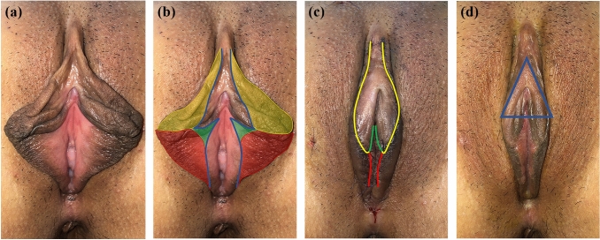
Fig. 3.
Trilobal labiaplasty. a Preoperative figure of a patient with bilateral labia minora hypertrophy and lateral clitoral folds. b The unstained area within the blue line was preserved tissue, the yellow area represents the clitoral hood reduction, the green area represents the wedge resection, and the red area represents the edge resection. c Three flaps were sutured together to form new labia minora. d A postoperative figure is shown. The structure in blue triangle was “Clitoral-labial triangle”

