Figure 1.
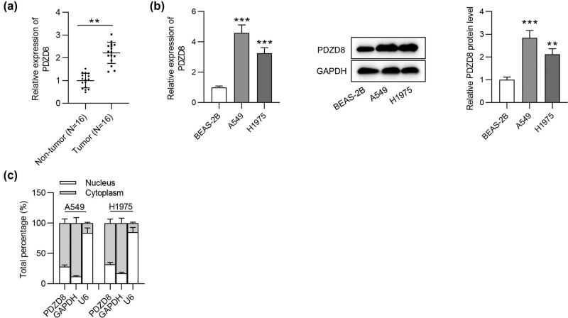
PDZD8 is highly expressed in LUAD tissues and cells. (a) PDZD8 expression in LUAD tissues (n = 16) was elevated compared with that in normal tissues (n = 16) as shown by RT-qPCR. **p < 0.01. (b) The mRNA and protein levels of PDZD8 in LUAD cells were increased as suggested by RT-qPCR and western blot analyses. **p < 0.01, ***p < 0.001 vs BEAS-2B group. (c) Subcellular fractionation assays revealed that PDZD8 was mainly distributed in cytoplasm of LUAD cells. The data are presented as the mean value ± standard deviation. Student’s t test was used to compare differences between LUAD tissues and normal tissues. One-way analysis of variance followed by Tukey’s post hoc analysis was used to compare differences among groups.

