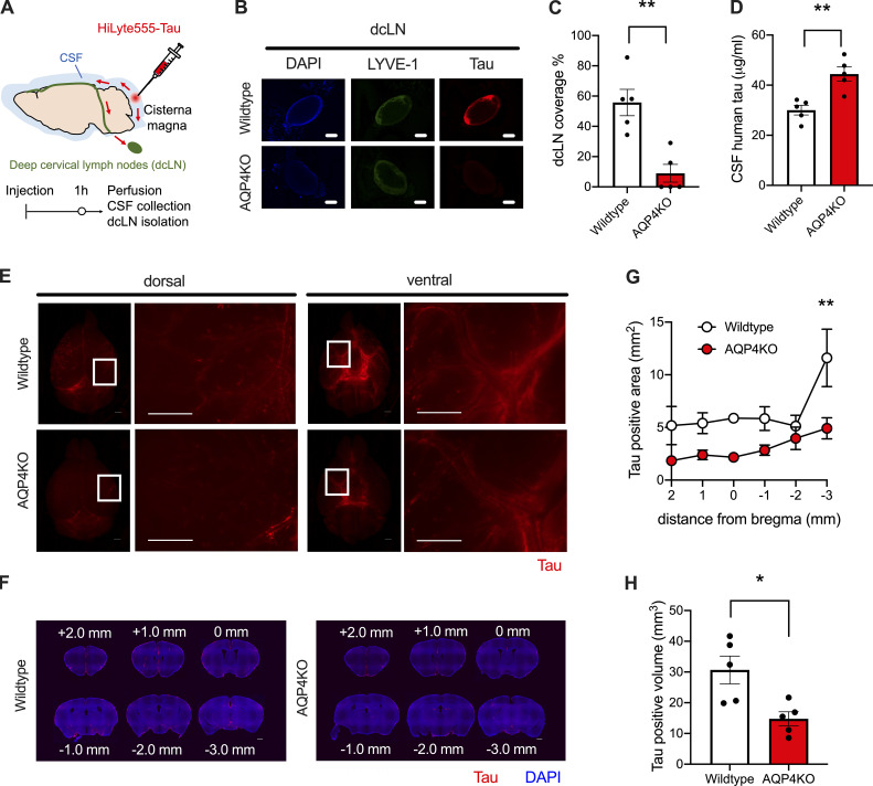
Figure 2.
Tau clearance from CSF to dcLNs and influx into the brain occur in an AQP4-dependent manner. (A) At 1 h from intracisternal injection of HiLyte 555–labeled tau, CSF, brains, and dcLNs were analyzed. (B) Representative images of tau accumulation (red) in LYVE-1–positive dcLNs (pseudocolored green) with DAPI counterstaining (blue). Scale bars, 1 mm. (C) Percent area covered by tau in dcLNs. Unpaired two-tailed test; **, P < 0.01. n = 5/group. (D) CSF tau levels. Unpaired two-tailed test; **, P < 0.01. n = 5/group. (E) Representative images of tau accumulation (red). Scale bars, 1 mm. (F) Representative brain sections showing tau (red) accumulation with DAPI staining (blue). Scale bars, 1 mm. (G) Tau-positive area (mm2). Two-way ANOVA with Bonferroni post-hoc analysis; **, P < 0.01. n = 5/group. (H) Average tau-positive volume (mm3). Unpaired two-tailed test; *, P < 0.05. n = 5/group.

