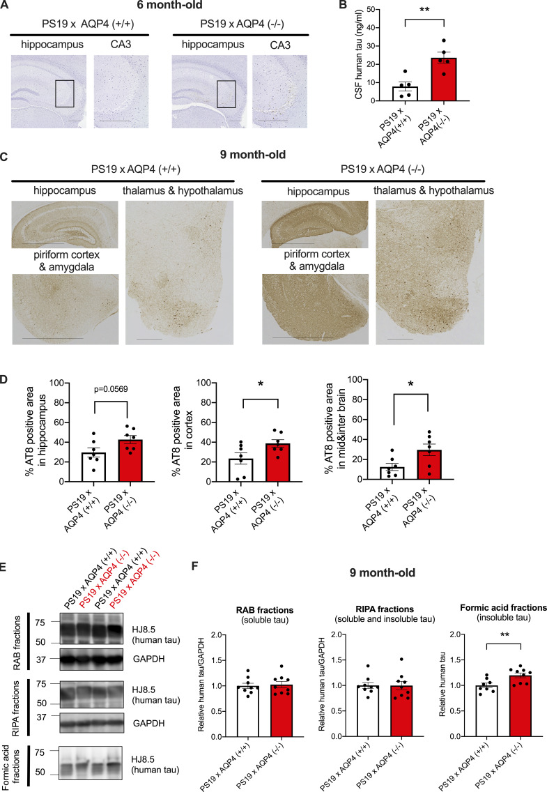
Figure 3.
AQP4 deficiency markedly exacerbates tau pathology in PS19 mice. (A) Representative images of AT8 staining in 6-month-old mice with hematoxylin staining. Scale bars, 300 μm. (B) CSF human tau levels in 6-month-old mice. Unpaired two-tailed t test; **, P < 0.01. n = 5/group. (C) Representative images of AT8 staining in 9-month-old mice. Scale bars, 1 mm for the hippocampus, piriform cortex, and amygdala and 300 μm for the thalamus and hypothalamus. (D) Quantification of the percentage of area covered by AT8 staining in 9-month-old mice. Unpaired two-tailed t test; *, P < 0.05. n = 7/group. (E) Representative immunoblots probing for human tau and GAPDH in RAB, RIPA, and formic acid fractions of 9-month-old PS19 × AQP4 (+/+) and PS19 × AQP4 (−/−) mice (in kD). (F) Quantification of immunoblot probing for human tau in RAB, RIPA, and formic acid fractions of 9-month-old PS19 × AQP4 (+/+) (n = 8 or 9) and PS19 × AQP4 (−/−) mice (n = 9). Unpaired two-tailed t test; **, P < 0.01. Source data are available for this figure: SourceData F3.

