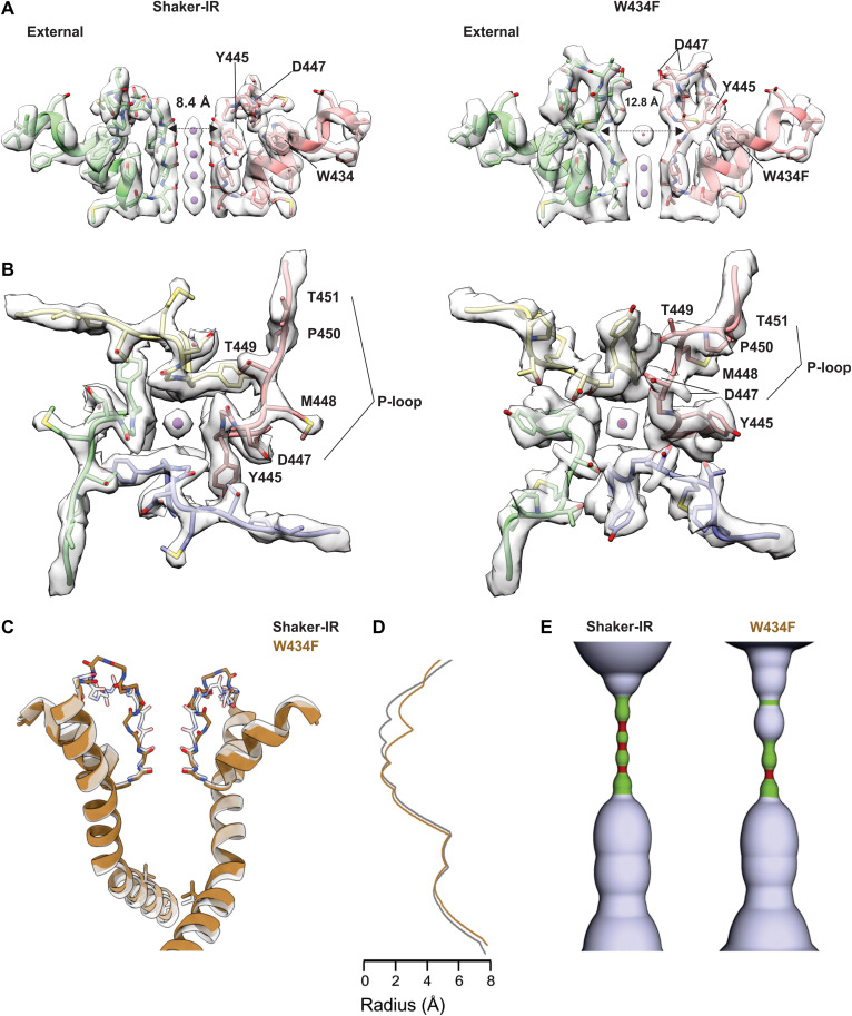
Fig. 2. Structure of the external pore of wild-type Shaker-IR and the W434F mutant.
(A) Side view of the selectivity filter of Shaker-IR (left) and W434F (right). The distance between Y445 CA is shown to highlight the dilation of the selectivity filter in W434F. (B) View of the pore domain of Shaker-IR (left) and W434F (right) viewed from an extracellular perspective. Residues experiencing a large movement are labeled to highlight structural reorientation of the outer pore domain in W434F. (C) Superposition of pore-lining regions for Shaker-IR and the W434F mutant. V478 is shown to mark the position of the internal S6 gate that prevents ion permeation across the internal pore when the Shaker Kv channel closes (51). (D) Plot of pore radius for Shaker-IR and the W434F mutant. (E) Hole diagrams illustrating the ion permeation pathways for Shaker-IR and W434F. Radii ≤1 Å are shown in red; radii >1 Å and ≤ 2 Å are shown in green, and radii larger than 2 Å are shown in light blue. The plot in (B) and the diagram in (C) are aligned with the protein model in (A).

