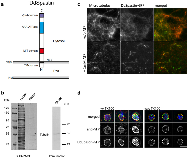
Figure 1.

Domain conservation and membrane orientation of DdSpastin. (a) Schematic of DdSpastin domains and membrane orientation by motif predictions using ELM 25, 26. See text for further descriptions; PNS, perinuclear space. (b) Immunoprecipitation using GFP-Trap Agarose beads showing tubulin-binding of DdSpastin-GFP. Proteins in the supernatant (lysate; corresponding to ~106 cells) and the GFP-Trap eluate (corresponding to 1 × 10⁷ cells) were separated by SDS-PAGE, and stained with Coomassie or evaluated by immunoblot staining with anti-β-tubulin; *, this particular band was analyzed by mass spectrometry resulting in a hit for α-tubulin (see table S1). (c) In vitro microtubule severing assay. Polymerized porcine brain tubulin and DdSpastin-GFP (green) were incubated with and without 1 mM ATP. The reaction mixture was fixed with formaldehyde on poly-L-lysine coated coverslips and stained with anti-α-tubulin (red). Green spots most likely represent DdSpastin-GFP clusters that have formed via hydrophobic interactions of the transmembrane domains. Bar, 5 µm. (d) Verification of membrane orientation using isolated nuclei from DdSpastin-GFP overexpression cells. Nuclei were fixed with and without Triton X-100 permeabilization. Merged images of three examples each and corresponding single channel images are shown. Bar, 2 μ
