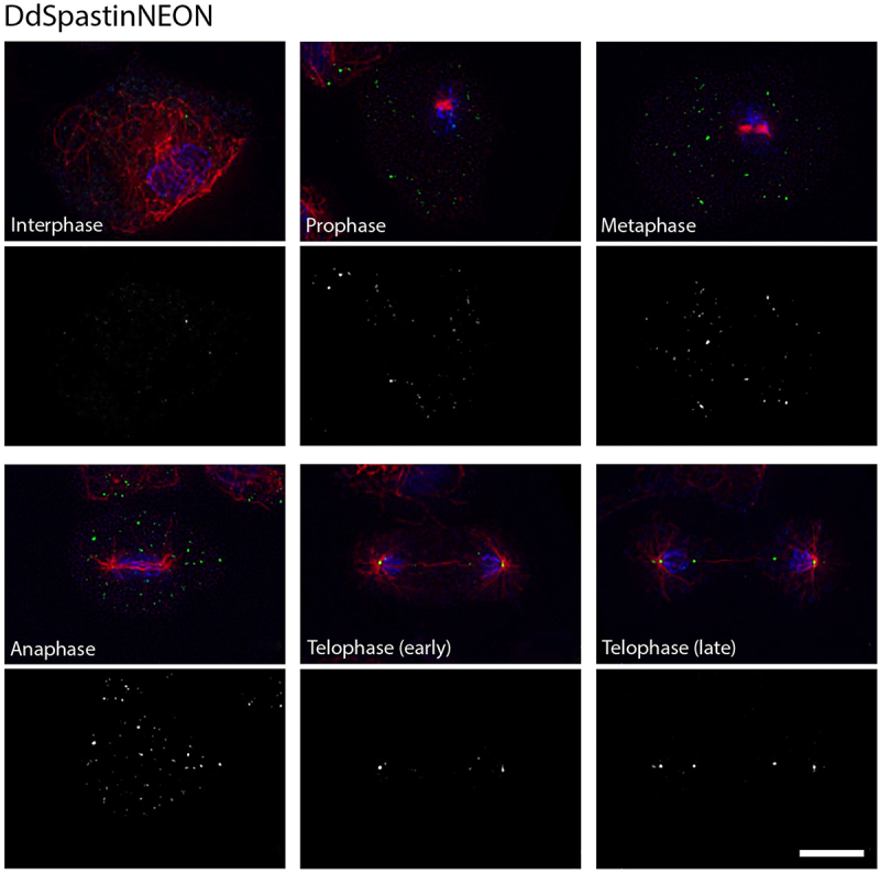
Figure 2.

Localization of DdSpastin-NEON (knock-in). Cells were fixed with glutaraldehyde and stained with DAPI(blue), and anti-α-tubulin (red). DdSpastin-NEON (green) accumulated at spindle Poles beginning in early telophase and in late telophase at the central spindle. The DdSpastin-NEON channel alone is shown below the merged images. A quantitative evaluation of all investigated cells is given in Table S2. Bar, 5 μm.
