Figure 3.

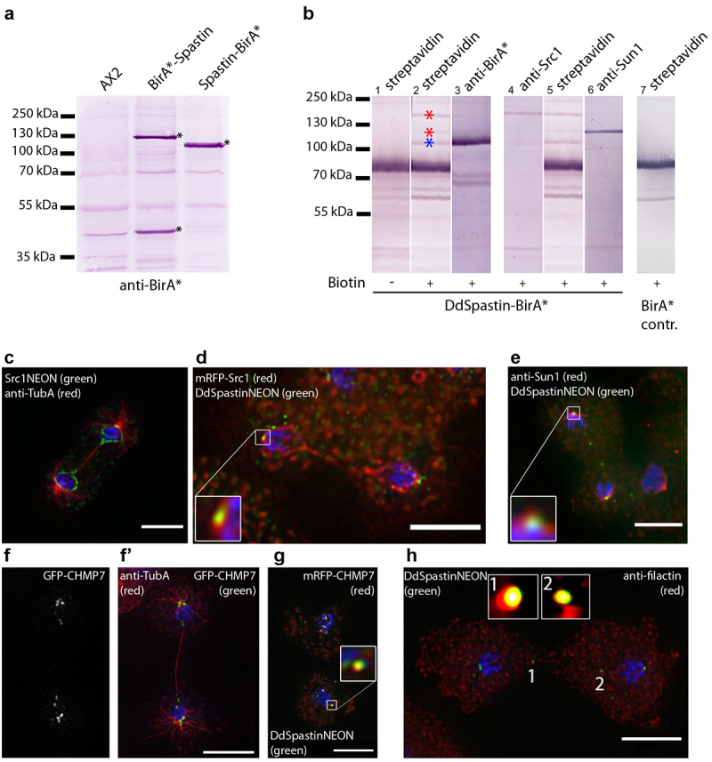
DdSpastin interactions and co-localizations. (a) Immunoblot of whole cell extracts of AX2 control cells, BirA*-DdSpastin cells and DdSpastin-BirA* cells stained with anti-BirA* antibodies. Fusion protein bands and BirA* with signal peptide are labeled with an asterisk. (b) BioID with nuclear extracts of DdSpastin-BirA* (lane 1–6) and negative control BirA* cells (lane 7). Western blots were stained with alkaline phosphate conjugated to the antibodies/protein stated on top. The interactors Src1 and Sun1 are labeled with red asterisks, DdSpastin-BirA* is labeled with a blue asterisk (lane 2). Lane 1 control w/o biotin incubation and lane 7 BirA* control show no specific bands at these positions. (c-h) Fluorescence microscopy of the strains stated in the figures. Cells were fixed with either glutaraldehyde (c-g) or methanol (h), and additionally labeled with DAPI (blue). Close-ups show co-localization (d, e, g, h). GFP-CHMP7 labeling is shown in a single channel because this protein has not been previously published (f). Bar, 5 μm.
