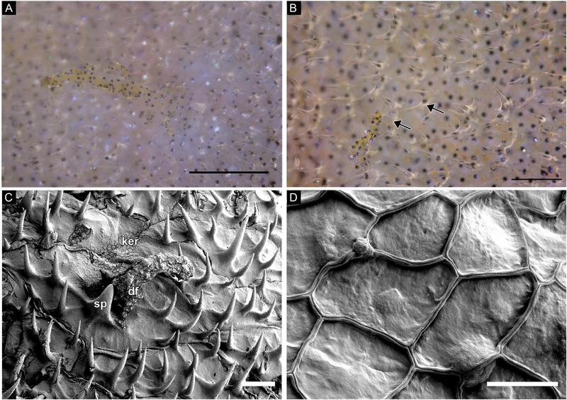
Figure 7:
(A-B) Freshly dead tissue; photographs of a branched dermal flaps covered with brown and yellow melanocytes and surrounded by smaller, transparent, forked spinules. (C) SEM image of a dermal flap surrounded by hook-like spinules. Squamous keratinocytes overlie the surface of the skin. Note the outlines of scales. (D) SEM image of the squamous keratinocytes. Abbreviations: dermal flap (df), keratinocytes (ker), spinule (sp). Scale bars: A-B, approx. 100μm; C, 100μm; D, 10μm.

