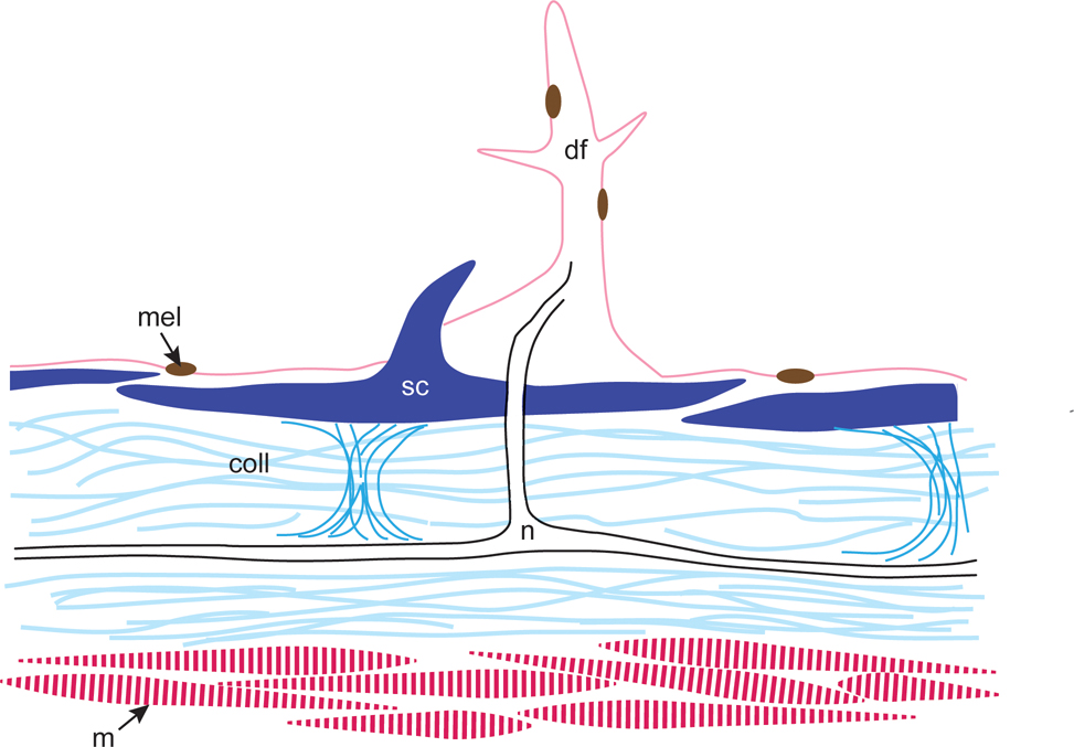
Figure 9:
Schematic diagram of a dermal flap in cross section showing skin elements. Scales are held in place with collagen; nerve fibers between collagen layers extend through an overlying scale to the core of the dermal flap. Light and medium blue, collagen (coll); pink, dermal flap (df), dark pink, muscle (m); brown, melanophore (mel); black, nerve tract (n); dark blue, scales (sc), center scale with spinule.

