. 2022 Feb 18;11:e71705. doi: 10.7554/eLife.71705

Figure 3. eIF2α is phosphorylated in ribosomal protein mutants via Xrp1 and PERK.

Panels A-J show single confocal planes from third instar wing imaginal discs. (A) Mosaic of RpS17+/- and RpS17+/+ cells. p-eIF2α levels were increased in RpS17+/- cells (see A’). (B) Mosaic of RpL27A+/- and RpL27A+/+ cells. p-eIF2α levels were increased in RpL27A+/- cells (see B’). (C) Clones of cells expressing Xrp1-RNAi in a RpS17+/- wing disc in white p-eIF2α levels were reduced by Xrp1 depletion (see C’). (D) Clones of cells expressing Xrp1-RNAi in a RpS17+/- wing disc in white. Translation rate was restored by Xrp1 depletion (see D’). (E) Clones of cells over-expressing PPP1R15 in a RpS17+/- wing disc in white. p-eIF2α levels were reduced by PPP1R15 over-expression (see E’). (F) Clones of cells over-expressing PPP1R15 in a RpS17+/- wing disc in white. Translation rate was restored by PPP1R15 over-expression (see F’). (G) Clones of cells expressing PERK-RNAi in an otherwise wild type wing disc in white. p-eIF2α levels were unaffected (see G’). Note that in this and some other panels mitotic cells are visible near the apical epithelial surface. Mitotic figures, which lack OPP incorporation, are labeled by the anti-p- eIF2α antibody from Thermofisher, but not by the anti-p- eIF2α antibody from Cell Signaling Technologies. (H) Clones of cells expressing PERK-RNAi in a RpS17+/- wing disc in whiite. p-eIF2α levels were reduced by PERK knockdown (see H’). (I) Clones of cells expressing PERK-RNAi in a RpS17+/- wing disc in white. Translation rate was restored by PERK knockdown (see I’). (J) Clones of cells expressing Gcn2-RNAi in a RpS17+/- wing disc in white. p-eIF2α levels were not reduced by Gcn2 knockdown (see J’). Further data relevant to this Figure are shown in Figure 3—figure supplement 1. Genotypes: A: p{hs:FLP}/+; RpS17 p{arm:LacZ} FRT80B/FRT80B, B: p{hs:FLP}/ p{hs:FLP}; RpL27A- p{arm:LacZ} FRT40/FRT40, C, D: p{hs:FLP}/+; RpS17, act> CD2> Gal4, UAS-GFP /UAS- RNAiXrp1, E,F: p{hs:FLP}/+; UAS-PPP1R15/+; RpS17, act> CD2> Gal4, UAS-GFP /+, G: p{hs:FLP}/+; UAS- RNAiPERK /+;act> CD2> Gal4, UAS-GFP /+, H, I: p{hs:FLP}/+; UAS- RNAiPERK /+; RpS17, act> CD2> Gal4, UAS-GFP /+, J: p{hs:FLP}/+; UAS- RNAiGcn2/+; RpS17, act> CD2> Gal4, UAS-GFP /+.

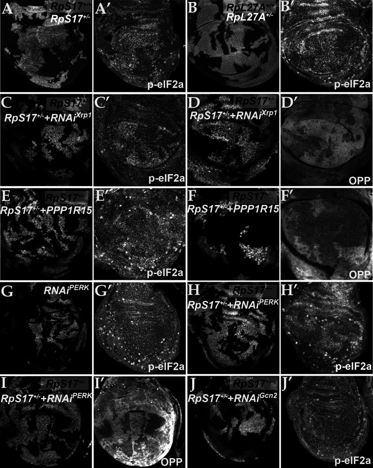
Figure 3.

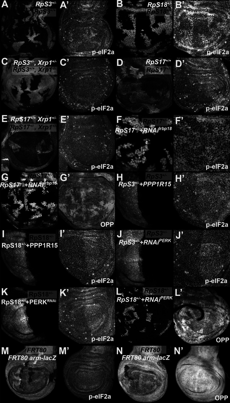
Figure 3—figure supplement 1. eIF2α phosphorylation in Rp+/- cell depends on Xrp1 and Irbp18.

Figure 3—figure supplement 1.

Panels A-L show single confocal planes from third instar wing imaginal discs. (A) Mosaic of RpS3+/- and RpS3+/+ cells. p-eIF2α levels were increased in RpS3+/- cells (see A’). (B) Mosaic of RpS18+/- and RpS18+/+ cells. p-eIF2α levels were increased in RpS18+/- cells (see B’). (C) Mosaic of RpS3+/-Xrp1+/-and RpS3+/+Xrp-/- cells. p-eIF2α levels were unaffected in RpS3+/- cells when Xrp1 was mutated (see C’). (D) Mosaic of RpS17+/- and RpS17+/+ cells (the latter brighter white, having two copies of β-gal transgene). p-eIF2α levels were increased in RpS17+/- cells (see D’). (E) Mosaic of RpS17+/- and RpS17+/+ cells in an Xrp1+/- disc, (RpS17+/+ brighter white, having two copies of β-gal transgene). p-eIF2α levels were unaffected in RpS17+/- cells (see E’). (F) Clones of cells expressing Irbp18 RNAi in a RpS17+/- wing disc in white. p-eIF2α levels were reduced by Irbp18 knock-down (see F’). (G) Clones of cells expressing Irbp18 RNAi in a RpS17+/- wing disc in white. Translation rate was restored by Irbp18 knock-down (see G’). (H) Cells over-expressing PPP1R15 in the posterior compartment of a RpS3+/- wing disc in white. p-eIF2α levels were reduced by PPP1R15 over-expression (see H’). (I). Cells over-expressing PPP1R15 in the posterior compartment of a RpS18+/- wing disc in white. p-eIF2α levels were reduced by PPP1R15 over-expression (see I’). (J) Cells expressing PERK RNAi in the posterior compartment of a RpS3+/- wing disc in white. p-eIF2α levels were reduced by PERK knock-down (see J’). (K). Cells expressing PERK RNAi in the posterior compartment of a RpS18+/- wing disc in white. p-eIF2α levels were reduced by PERK knock-down (see K’). (L) Clones of cells expressing PERK RNAi in a RpS18+/- wing disc in white. Translation rate was restored by PERK knock-down (see L’). (M) Neutral clones expressing or lacking LacZ expression did not affect p-eIF2α levels (see M’). (N) Neutral clones expressing or lacking LacZ expression did not affect OPP incorporation (see N’). Genotypes: A: p{hs:FLP}/ p{hs:FLP}; FRT82 RpS3 p{arm:LacZ} /FRT82B, B: p{hs:FLP}/ p{hs:FLP}; FRT42 RpS18 p{Ubi:GFP}/FRT42, C: p{hs:FLP}/ p{hs:FLP}; FRT82 RpS3 p{arm:LacZ} /FRT82B Xrp1M2-73, D: p{hs:FLP}/ p{hs:FLP}; RpS17 FRT80B/p{arm:LacZ} FRT80B, E: p{hs:FLP}/ p{hs:FLP}; RpS17 FRT80B/p{arm:LacZ} FRT80B Xrp1M2–73, F-G: p{hs:FLP}/+; RpS17, act> CD2> Gal4, UAS-GFP /UAS- RNAiIrbp18, H: en-GAL4, UAS-GFP /UAS-PPP1R15; FRT82 RpS3/+, I: RpS18-,en-GAL4, UAS-GFP /UAS-PPP1R15, J: en-GAL4, UAS-GFP / UAS- RNAiPERK; FRT82 RpS3/+, K: RpS18-,en-GAL4, UAS-GFP /UAS- RNAiPERK, L: p{hs:FLP}/+; UAS- RNAiPERK / RpS18-; act> CD2> Gal4, UAS-His-RFP/+, M, N: p{hs:FLP}/ p{hs:FLP}; FRT80B/p{arm:LacZ} FRT80B.