Figure 3.

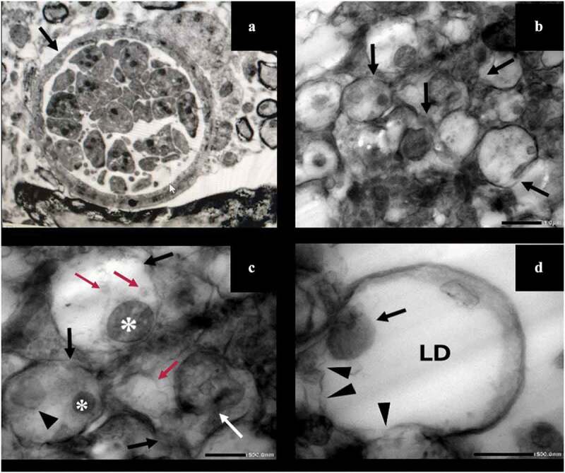
TEM of T. gondii cysts in brain homogenate of the avirulent Me49 strain-infected non-treated subgroup (Ib) (A) and the infected aluvia-treated one (IIa3) (B-D). (A) T. gondii cyst revealing an intact well circumscribed cyst wall (arrow) containing multiple bradyzoites with intact cell membranes (x 3000). (B) Multiple T. gondii bradyzoites showing disrupted plasma membranes (arrows) (x 5000). (C) Bradyzoites demonstrating marked discontinuity of their plasma membranes (black arrows), lipid droplets of varying sizes and densities (red arrows), swollen mitochondria (asterisks), dilated endoplasmic reticulum (arrow head) and a localized disruption of the nuclear envelope (white arrow) (x 15,000). (D) A bradyzoite possessing a swollen mitochondrion (arrow) and a large electron-lucent lipid droplet (LD) with evident disintegration of its plasma membrane (arrow heads) (x 20,000).
