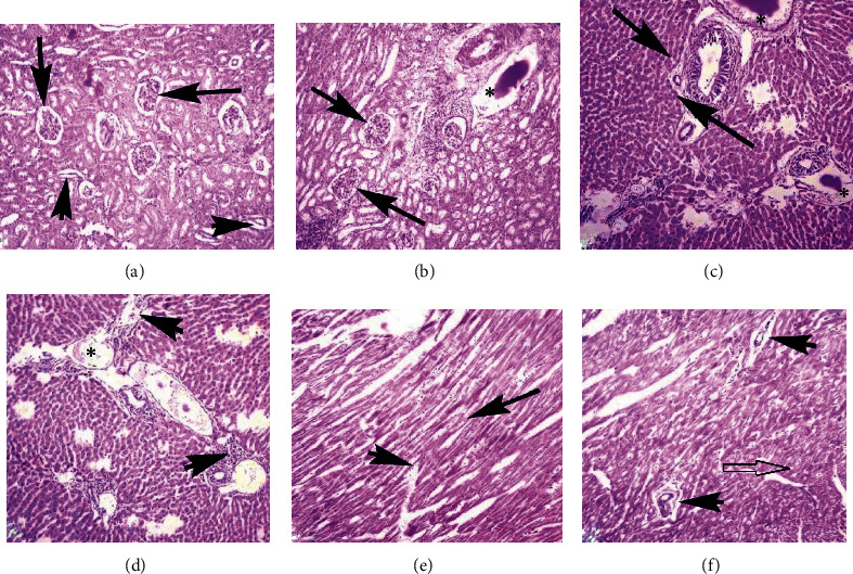
Figure 7.

Photomicrograph showing necrosis of renal tubular epithelium, increased urinary space (arrows), degeneration of renal tubules and disorganization of glomeruli (arrowheads), and edema (∗) in the kidneys (a and b); degeneration and pyknosis of hepatocyte, edema (∗), fatty change (arrow), and inflammatory materials (arrowheads) in various sections of the liver (c and d); and coagulative necrosis (arrow), inflammatory exudate, disruption of cardiac muscles (arrowheads), and vacuolar degeneration (empty arrow) in heart sections of the rabbits (e and f) at day 10 of exposure (400x, H&E stain).
