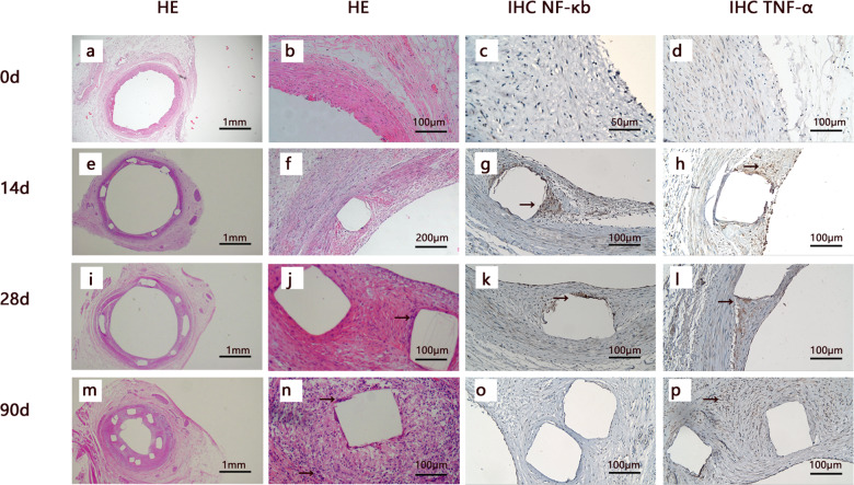
Fig. 1.
Histology and immunohistochemistry of porcine coronary arteries implanted with PLLA-BVS follow-up to 90 d. The images were shown by HE staining (from left to right, first and second columns) and immunohistochemistry staining (third and fourth columns) of the coronary arteries after implantation for 0 d (a–d), 14 d (e–h), 28 d (i–l), 90 d (m–p), respectively. White squares in all pictures indicate components of PLLA stent, macrophages were indicated by black arrows at 28 and 90th day (j, n) in HE stain image, and positive immunohistochemistry staining of NF-κb/TNF-α were indicated by black arrows at 14 d, 28 d and 90 d (g, h, k, l, p).

