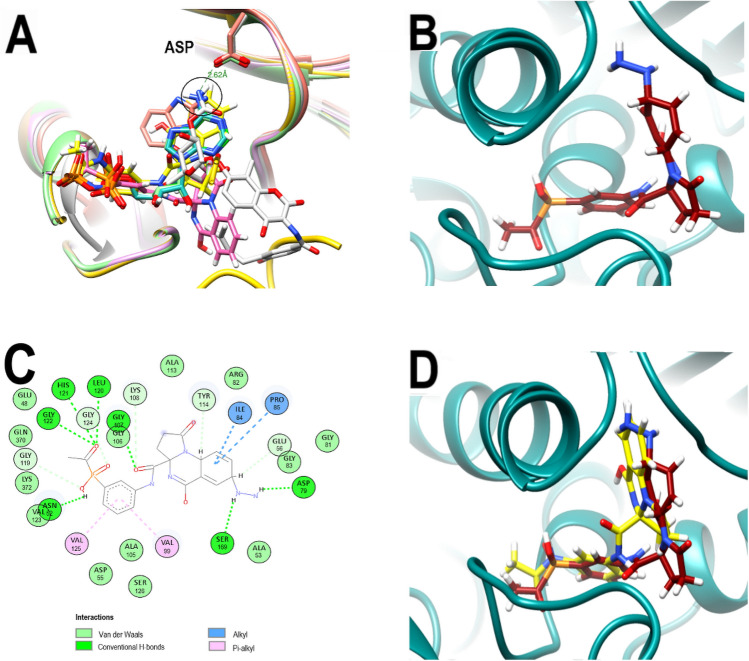
Figure 4.
Comparative poses of co-crystallized ligands of diverse GyrBs and analysis of the best-predicted docking pose of PQPNN. (A) Computationally performed overlay of PQd and co-crystallized ligand-with GyrBs: ADP (cyan), ATP (light green) AX7 (orange), novobiocin (white), ANP (green), PQd (yellow) and PQP (magenta). The black circle highlights the overlapping nitrogen of the co-crystallized ligands. (B) 3D representation of the 3ZKBL-PQPNN complex. (C) 2D interaction diagram of the 3ZKBL-PQPNN complex. (D) 3D representation of PQPNN-3ZKBL complex superimposed with PQd (yellow backbone).

