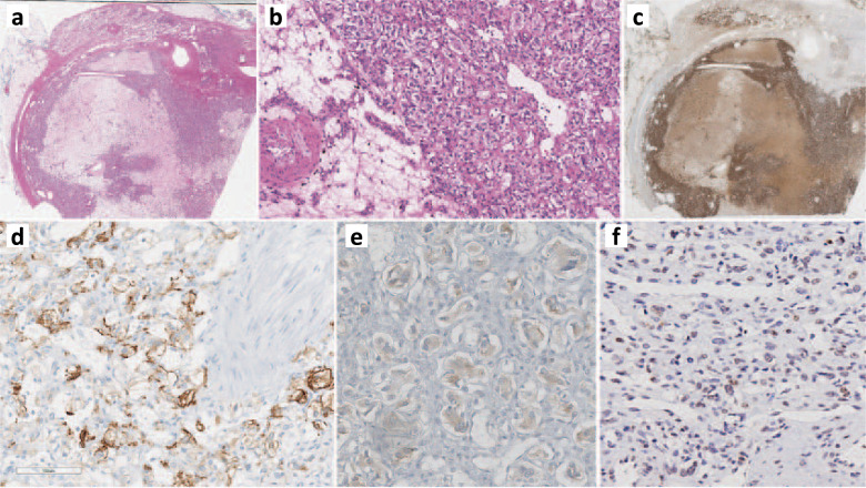
Fig. 1. Morphological and immunohistochemical findings of pheochromocytoma.
Whole scanned images: The adrenalectomy specimen (hematoxylin and eosin) shows an encapsulated pheochromocytoma with clear cell change, variable fibrohyaline, and myxoid stroma rich in microvasculature (a, b). The tumor cells are diffusely positive for tyrosine hydroxylase (the rate limiting enzyme in catecholamine synthesis) (c). The tumor is positive for carbonic anhydrase IX (d). Alpha-inhibin shows variable weak reactivity in the tumor cells (e). pVHL shows variable loss or significantly reduced staining intensity in the tumor cells (f).

