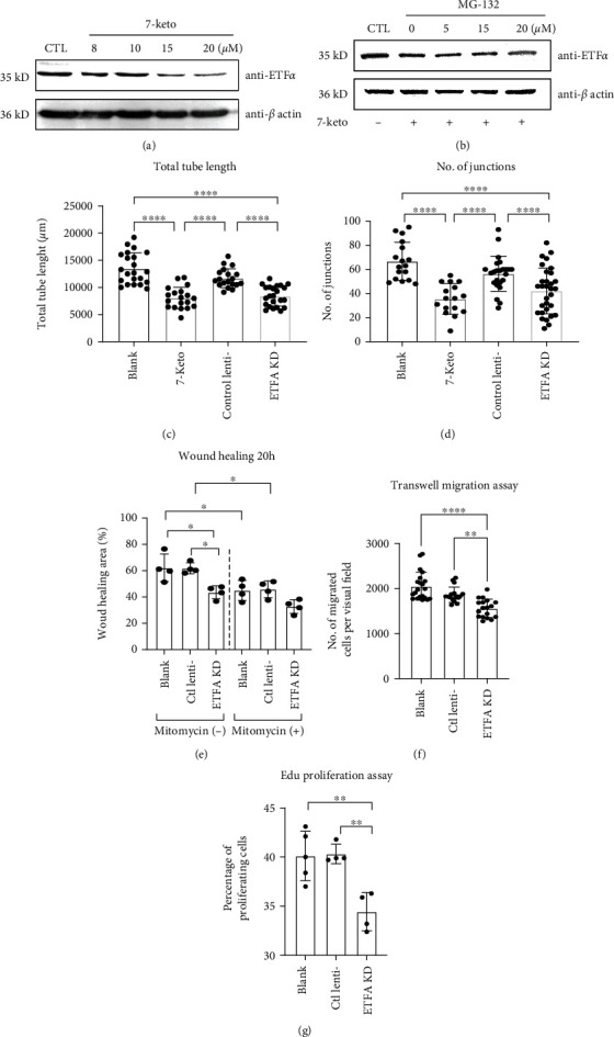
Figure 6.

The role of ETFα in vessel sprouting in vitro. (a) Representative immunoblot of ETFα in HUVECs stimulated by 7-keto cholesterol (8, 10, 15, and 20 μM). GAPDH was used as loading control; (b) representative immunoblot of ETFα in HUVECs stimulated by 20 μM 7-keto cholesterol or 7-keto with different doses of MG-132 (5, 15, and 20 nM). β-Actin was used as loading control; (c) quantitative analysis of the total tube length and (d) number of junctions in the tube formation assay (one-way ANNOVA, ∗P < 0.01, ∗∗P < 0.001, n = 20); (e) quantitative analysis of the wound closure area in HUVECs with the indicated treatments at different time points (one-way ANNOVA, ∗P < 0.01, n = 4); (f) quantitative analysis of transwell migration assay in HUVECs with the indicated treatments (one-way ANNOVA, ∗∗P < 0.001, ∗∗∗∗P < 0.00001, n = 15); (g) quantitative analysis of Edu proliferation assay in HUVECs with the indicated treatments (one-way ANNOVA, ∗∗P < 0.001, n = 4).
