
Figure 2.
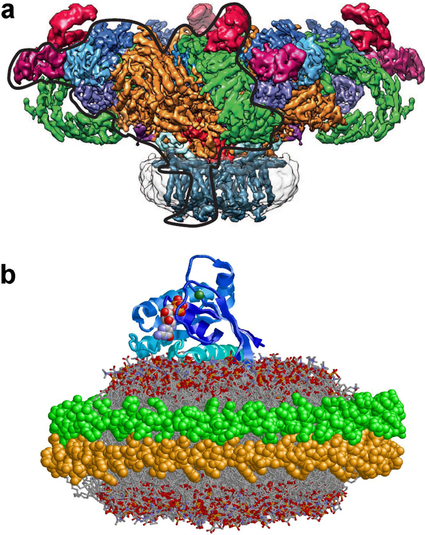
Examples of membrane protein structure in Nanodiscs. (A) The structure of the ryanodine receptor in Nanodisc is resolved to ~6.1 Å using cryo-electron microscopy. The Nanodisc is shown as a light gray envelope with 24 transmembrane helices forming a square structure similar to that of the incorporated voltage-gated sodium channel Nav (reproduced with permission from21). (B) The structure of KRas4b bound to the surface of a Nanodisc as determined by NMR spectroscopy 38 (2MSC.pdb file from the Protein Data Bank).