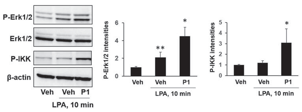
FIGURE 2.

Peptide P1 promotes LPA-induced phosphorylation of Erk1/2 and I-κB kinase (IKK) in MLE12 cells. MLE12 cells were transfected with P1 plasmid for 48 h, and then cells were treated with LPA (1 μM) for additional 10 min. Phospho-Erk1/2, Erk1/2, p-IKK, and β-actin levels were examined by immunoblotting. Blots were analyzed with ImageJ. n = 3, *p < .05, compared to Veh; **p < .01, compared to Veh. Shown are representative blots from three independent experiments. LPA, lysophosphatidic acid receptor
