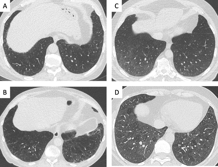
Fig. 6.
A–D. Collateral (smoking-related) findings in screening LDCT:interstitial lung abnormalities with varying extent and morphology.Axial CT image at the level of mid-lower chest showing different patterns of interstitial lung abnormalities with varying severity: A minor reticulation in right lateral sulcus; B reticulation with signs of bronchiolar traction in the lower lobes; C ground-glass opacity with mild extent in the lower lobes; D ground-glass opacity with extensive distribution in the lower lobes, associated with minimal areas of parenchymal sparing with lobular distribution. These findings variably represent smoking related disease, with either reversible or irreversible behaviour worth of multidisciplinary discussion

