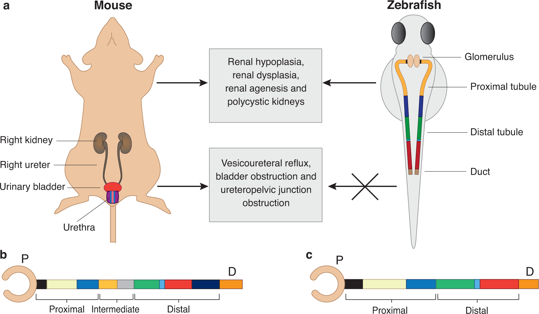
Figure 2. In-vivo and in-vitro models to study CAKUT.

A. Schematics of mouse (left) and zebrafish (right) urinary system. Mammals have two symmetrical kidneys, each of which are connected to the urinary bladder through a collecting ducting (ureter). The mammalian kidney is composed of millions of kidney structural units known as nephrons. The zebrafish urinary system is comprised of two functional pronephric tubules but share structural and functional similarity with mammalian urinary system. The absence of lower urinary tract organs is a limitation of the zebrafish model.
B. Schematic of the mammalian kidney structural unit is represented as a straightened nephron tube. The glomerulus capsule (blood filter) is drawn as a pink cup-like sac structure. The tubule can be subdivided in to three segments including proximal, intermediate and distal tubule. The proximal tubule consists of the neck (black), proximal convoluted tubule (yellow) and proximal straight tubule (blue). The intermediate tubule consists of descending thin limb (light orange) and ascending thin limb (gray). The distal tubule consists of thick ascending limb (green), macula densa (light blue), distal convoluted tubule (red) and connecting tubule (indigo). The last segment of nephron consists of collecting duct (orange).
C. The zebrafish pronephros is drawn as straight tubule. The glomerulus capsule is represented as pink cup-like sac structure. The zebrafish tubule lacks the intermediate segment and contains only the proximal segment (neck, proximal convoluted tubule and proximal straight tubule; black, yellow and blue, respectively) and distal portion (distal early, corpuscle of stannius and distal late tubule; green, light blue and red, respectively). The last segment of nephron is the duct (orange, also known as cloaca).
